Fiber Photometry Solutions

The Technique
Fiber photometry is a neuroimaging technique that monitors neuronal activity in freely-moving animals. This technique utilises genetically encoded fluorescent indicators (e.g. GCaMP, dLight, GRAB-Ach, RCaMP, jRGECO1) expressed in target brain regions. These indicators fluorescence only when bound to melocules such as calcium, dopamine or acetylcholine, etc. By implanting an optic fiber in the brain, we can excite and collect the result fluroescence emission reporting real-time molecular dynamics during complex behaviors.
Thus, unlike Doric Miniscopes, which resolve single-cell activity, fiber photometry records population signals (i.e bulk fluorescence from labelled neurons). This approach is ideal for circuit-level studies, espeically ones touching deep brain regions, like amgdala, hypothalamus, nucleus accumbens, pons, etc. By implanting multiple fibers in different interconnected nuclei, scientist can investigate changes in neural circuits during behavior, learning, memory, addiction, feeding, anxiety.
The fiber photometry approach provides several advantages over miniscope:
- Minimally invasive (especially for deep brain regions)
- Optimized for multi-animal & multi-site experiments
- Cost-effective
Fiber Photometry Systems
Doric Lenses Inc. is a recognized leader in advanced fiber photometry solutions for behaving animals, driving innovation in this rapidly evolving field.
All Doric Fiber Photometry system are complete, plug-and-play solution. We provide everything from cannula, fiber-optic patchs, rotary joint, optical cube, light sources, console, data acquisiiton software and analyisis software.
From long-term freely-moving animals, optogenetics capabilities, wireless, multi-fiber, etc. each system was carefully engineered to address the specific experimental needs & challenges.
The three main types of Fibers Photometry Systems are split into categories based on the type of detector:
- BASIC: photodetector with high sensitivity and high temporal resolution (FMC, RFMC, & WiFP)
- BUNDLE-IMAGING: CMOS camera images multiple fibers simultaneously (BFMC, BFTO, RBFMC)
- LIFETIME: fast photodetector measures changes in fluorescence lifetime at picosecond level (FluoPulse™)


Featured Products

FluoPulse™
Beyond intensity - measure lifetime.
Quantify tonic, relative baseline & slow-state dynamics via picosecond changes in fluorescence lifetime, in freely-moving animals.

Wireless System
A cable-free solution for experiments requiring multiple, interacting animals. Ideal for recording / manipulating the neural populations underlying social behaviors.

Rotary Bundle-imaging system
A high-power, multi-color light source for most available opsins on the market.
Fiber Photometry
This side-by-side comparison highlights differences in channel configuration, maximum number of site/animal(s), dual-color & optogenetics compatibility, and advanced features across seven distinct fiber photometry systems.
LEGEND:
- Iso. = isosbestic
- Opto = optogenetics
- Ephys = Electrophysiology
- EEG = elctroencephalogram

|

|

|

|

|

|

|
|
|
Ideal for 1-2 animals and/or 1-2 brain regions. Dual-colour photometry that combines readily with far-red optogenetics. |
For a single freely-moving animal, 1-2 site fiber photometry recording over long time period (hours-days). No rotational artifacts! |
Cable-free fiber photometry for freely-moving animals. Ideal for multiple interacting animals in the same cage. |
Affordable, high-throughput fiber photometry for multi-site / multi-animal experiments. |
High-throughput fiber photometry for multi-site / multi-animal experiments. Includes independent optogenetics per fiber. |
Ideal for single animal with three+ sites fiber photometry recording over long time periods (hours-days), without motion artifacts. |
Ideal for tonic and relative changes in baseline, in freely-moving animals. Longitudinal studies. |
|
|
MULTI-COLOR
|
|
|
|
|
|
|
|
|
MULTI-FIBER
|
|
|
|
|
|
|
|
|
ISOSBESTIC
|
Flexible: 405, 415, 425 & 440nm |
Fixed: 405, 415, 425 or 440nm |
Fixed: 405 or 415 nm |
Fixed: 405, 415, 425 or 440nm |
Fixed: 405, 415, 425 or 440nm |
Flexible: 405, 415, 425 & 440nm |
N/A |
|
Opto in DIFFERENT fiber: 470 nm, 520, 560nm, 590 nm, 638 nm |
N/A |
Opto in DIFFERENT fiber: 470 nm, 520, 560nm, 590 nm, 638 nm |
|
|
|
|
|
NO ROTATION ARTIFACTS
|
✓ |
✓ |
✓ |
✓ |
|||
|
|
|
Interleave |
Interleave |
Interleave |
Interleave |
|
|
|
|
|
|
|
1 - 25 Hz |
|
|
Rotary Joint: add Ephys / EEG |
|
Rotary Joint: add Ephys / EEG |
Rotary Joint: add Ephys / EEG |
|
Rotary Joint: add Ephys / EEG |
||
|
|
|
|
|
|
|


COMMON QUESTIONS:
What are the rotary systems?
What are the rotary systems?
These two types of light sources differ in terms of emitter size, maximum intensity, maximum illumniation area and recommend fiber diameter and NA.
LEDs
LEDs are among the most commonly used light sources in optogenetics, suitable for experiments stimulating large regions-of-interest (with 400 µm - 1mm diameter) at moderate light intensities. Their large emitter, inherent safety, reliability, and affordability make them a preferred option across a variety of experimental designs.
At Doric Lenses, we offer a wide range of CLED wavelengths in the near-UV (350-400 nm), visible (400-700 nm) and near IR range (700-1100 nm) for standard optogenetic applications.
Laser Diodes
Laser diodes are high-powered light sources with small emitter ideal for optogenetic experiments requiring high intensity of light in small regions-of-interest (< 200um).
Their narrow beam profile and monochromatic light output make lasers ideal for illumination through small-diameter patch cords (typically 50–200 µm) and are a great solution when precise spatial targeting is required.
What's unique about the LISER™ light source?
What's unique about the LISER™ light source?
LISER™ (Laser-Induced Spontaneous Emission of Radiation) is an advanced light source providing high-intensity, broad-spectrum illumination. The system uses a laser pumped Ce:YAG (cerium-doped yttrium aluminum garnet) crystal, which emits a continuous spectrum from yellow to red (500–650 nm).
The LISER™ provides exceptionally high optical power (> 85mW full spectrum in a 200 um NA 0.57 fiber) in a spectral range that is difficult to achieve with standard LEDs or laser diodes, particularly around 590 nm, optimal for activating inhibitory opsins such as NpHR and Jaws. In addition to its broad-spectrum output, LISER™ includes an integrated blue excitation source (laser or LED), making it a versatile, all-in-one solution capable of supporting both excitatory and inhibitory optogenetics simultaneously.
The LISER also includes its own internal drivers, allowing independent control of two wavelength channels in the same brain region (e.g. 450 nm + 590 nm).
Key Features:
- Full-Spectrum Coverage: Simultaneously delivers blue excitation plus tunable yellow-red wavelengths using interchangeable optical filters (e.g., 525, 559, 582, 593, 612 nm).
- High-Power: Ideal for experiments requiring intense illumination, including bilateral, and/or large-area stimulation.
- Supports Large Core Patch Cords for wide brain region illumination: Optimized for 200 - 400 µm core diameters, enabling uniform wide-area stimulation.
What is optimal light source for multi-animal experiments?
What is optimal light source for multi-animal experiments?
For multi-animal or multi-site optogenetics experiments, DazzLED is the optimal solution. This light source can be combined with the DazzLED Fiber Converter and Bundle Branching Fiber Optic Patch Cords to deliver high-power stimulation across many fibers simultaneously.
This configuration enables scalable, high-throughput photostimulation of up to 20 fibers with 400 µm diameter cores or up to 100 fibers with 200 µm diameter cores, making it well suited for parallel optogenetic experiments, multi-region stimulation, and large cohort studies.
Can light sources be triggered by TTL?
Can light sources be triggered by TTL?
To operate Doric LED and Laser diode modules, a dedicated light source driver is required. The driver can either come as a standalone device, or can integrate the ligth source (fiber-light sources, FLS).
- Integrated Driver Units (FLS): In this setup, each driver channel has a built-in LED or laser, offering a compact and user-friendly solution.
- Modular Driver Setup (Driver): The LED/laser remains separate from the driver, provides flexibility to switch between different wavelengths.
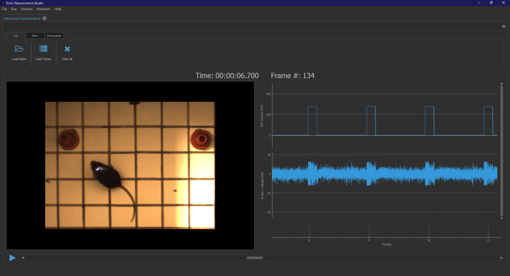
The driver can be operated in 1) standalone, 2) external modes (Analog or TTL), or 3) program sequences using free Doric Neuroscience Studio (DNS) software. In DNS, users can define a wide range of light stimulation patterns including continuous wave output, square pulse trains, sine waveforms, and cutoms waveforms.
Watch the tutorial video for step-by-step guidance on using LED drivers.
All light source drivers can be triggered by external digital signals, unlocking the ability to do close-loop experiments, where behaviors (lever press, nose poke, beam break, etc.) trigger the optogenetic stimulation. As such, DNS support complex triggering modes to pause, restart, and continue a stimulation pattern, as described in the following tutorial video.
NOTE:
- LED drivers are only compatible with LEDs, and LD drivers only with lasers.
- LISER come with integrated driver.
Basic Fiber Photometry System
All Basic Fiber Photometry Systems are fitted with a high-sensitivity Doric photodetector. This detector can be driven at high sampling rates and can be run in either lock-in mode (frequency-based division) or interleaved mode (time-based division) using live demodulation/deinterleave algorithms in (free) Doric Neuroscience Studio software.
The other primary difference between basic and bundle-imaging system is that one optical cube is required per optical fiber, so the price scales according to the number of sites/animals run simultaneously.
All Basic systems now come in integrated models where most components, such as the LEDs, LED Driver, optical components, detectors and even rotary joint are combined into a small compact form factor. Different systems have different levels of integration.

Fluorescence Mini Cubes (FMC)
In the latest generations, the photodetector is integrated directly within the fluorescence mini cube providing higher signal-to-noise ratio compared to non-integrated version. One FMC records a GFP-based and/or RFP-based signal from a single optical fiber. However, it is possible to combine multiple cubes/headstages to record from more than one mouse or brain site (up to 4-8*) when using the Neuroscience Console 500.
Fluorescence Mini Cube (FMC) now come with a flexible isosbestic point, such that the integrated isosbestic LED can be swapped between: 405, 415, 425 and 440 nm LEDs. In addition, both the 1-color and 2-color configuraitons are compatible with simultaneous red-shifted optogenetics, without crosstalk.
In addition, the Basic system can be combined with pigtailed rotary joints (FRJ_1x1_PT, FRJ_2x2_PT & AFRJ_2x2_PT) for long-term, freely-moving experiments.
With decade-long validation, the basic fiber photometry system is ideal to record GFP, RFP and isosbestic signals from 1-2 sites/animals, with the possibility to expand later on.
Rotary Basic Fiber Photometry
In Rotary Fiber Photometry Mini-Cube (RFMC), up to two fluorescence mini cube(s) are built-on the rotary joints itself. This unique design abolishes rotational artifacts in the signal and minimizes the number of connections before the photodetector. Therefore, RFMC system boasts a higher quality signal compared to basic FMC system paired with an external rotary joint, and is ideal for experiments lasting several hours or even days.
The rotary joint also contains a hollow channel that can be used for fluid delivery, electrophysiology/EEG, or optogenetics.


Wireless Fiber Photometry
The Wireless Fiber Photometry System (WiFP) completely circumvents tangling issues by eliminating the need for fiber optic cables fixed to the animal's head.
Moreover, this is the only solution to record from multiple freely-moving animals in the same cage, or for behaviors disrupted by tethered system. Currently, only 1-color + isosbestic option is availalble. Moreover, the WiFP is currently the only wireless fiber photometry solution available on the market with an isosbestic control signal.
Multiple headstages can be run at the same time, thus this system is ideal to study neural population underpinning social behaviors.
Bundle-Imaging Fiber Photometry Systems
Unlike the Basic Fiber Photometry Systems, which require one mini cube per site/animal, the Bundle-imaging Fiber Photometry systems use a CMOS camera to image the entire fiber bundle simultaneously. Bundle-Imaging Fiber Photometry System are ideal when recording 1- or 2-colors from multiple animals or brain regions (1-19 fibers), all at a reasonable price.
All bundle-imaging system are available with 2-color configurations (plus isosbestic) and the three excitations are interleaved in time (i.e the lock-in mode is not available). All three LEDs excite the entire fiber bundle (no independent power adjustement), and the photometry signal is then sampled at 20-100 Hz depending on the field-of-view of the camera and the number of fibers imaged at the same time.

Bundle-imaging Fluorescence Mini Cube (BFMC)
The BFMC-G3 is fully integrated, plug & play design (with CMOS camera, LEDs, LED Driver & console in a single device), greatly simplifying the set-up & troubleshooting. However, this system is not compatible with simultaneous optogenetics in the same fiber as fiber photometry measure.
Bundle-imaging with Targeted Optogenetics (BFTO)
The BFTO system is specifically designed for dual-colour, multi-site fiber photometry with targeted optogenetics. The term "targeted" refers to independent, multi-site optogenetic control, were multiple light sources are connected to different optic fibers within the bundle of fibers.
The BFTO is particularly well suited for circuit neuroscience because it enables simultaneous recording from multiple brain regions while delivering targeted optogenetic stimulation to specific nodes within the network. This combination can measure how activity propagates across interconnected circuits, determine causal relationships between regions, and dissect the functional role of defined pathways during behavior—all within the same experiment.
Moreover, the targeted optogenetics approach is also ideal for multi-animal, close-loop optogenetics, where each animal trigger the stimulation according to their unique behavior state.
With dual-color, multi-site and optogenetic capability, this system provides the greatest flexibility for common neuroscience experiments.


Rotary Bundle-imaging Fluorescence Mini Cube (RBFMC)
The RBFMC is the only rotary joint solution for multi-site fiber photometry experiments (1-color or 2-color + isosbestic) and can even be combine with electrophysiology/EEG.
This solution is designed for single, freely-moving animal and for experiments lasting several hours/days. The CMOS detector is integrated directly on the rotating joint ABOLISHING rotation-related artifacts. In addition, the 2025 version now comes with a flexible isosbestic design, to support switching between 405, 415, 425 and 440 nm for different biosensors.
The RBFMC also includes two different optogenetic capabilities: (1) a configuration for simultaneous GCaMP and red opsin in all fibers (simultaneously) and/or (2) and upgrade to fit with external laser diode on the rotary joint rotor, for independent optogenetic stimulation on a non-fiber photometry site.
The RBFMC system comes by default with integrated electrical connectors for Intan, neuropixel other electrophysiological recordings. This RBFMC system this ideal solution to combine multi-site fiber photometry and electrical measures in the same freely-moving animals.
* Simultaneous optogenetic on the fiber photomery sites is only available for the 1-color configuration; For 2-color configuration optogenetics can be added, but on a non-photometry site.
Fluorescence Lifetime Fiber Photometry (FLiP)
The latest innovation in fiber photometry! Fluorescence lifetime fiber photometry measures picosecond changes in the fluorescence lifetime of indicators rather than signal intensity, providing a readout that is largely independent of biosensor experession level, motion artifacts, photobleaching, varying excitation power.

FluoPulse™ System - Fluorescence lifetime fiber photometry
This approach is particularly ideal to quantify tonic, relatives baseline changes & slow-state dynamics of neurotransmitter, metabolites or kinases in freely-behaving animals. Fluorescence lifetime measures are also optimized for longitudinal studies, reducing variation between and within animals.
The FluoPulse system supports up to two channels simultaneously, either: dual-site, dual-animal or dual-color.
NOTE: The FLiP method requires special biosensors that have meanginful change in lifetime < 100 picoseconds. So classic biosensor like GCaMP are not compatible. For a full list of fluroescence lifetime biosensors (and those already validated with our system) check out the Biosensor Tab HERE.
What is fluorescence lifetime?
What is fluorescence lifetime?
Fluorescence lifetime is the average time a fluorophore remains in its excited state before emitting a photon, typically on the nanosecond timescale. It is an intrinsic property of the fluorophore that depends on its local molecular environment rather than its concentration or excitation intensity.
How is fluorescence lifetime measured?
How is fluorescence lifetime measured?
The Doric FluoPulse system uses a novel Pulse Sampling methedology to measure fluorescence lifetime. Unlike other single-photon counting (TCSPC) approaches, collects mutliple photons at each measurement.
Are special types of biosensors required to measure fluorescence lifetime?
Are special types of biosensors required to measure fluorescence lifetime?
Yes. To measure fluorescence lifetime, you must express specially engineered biosensors that have measurable change in fluorescence lifetime > 50 ps.
Classic intensity biosensors, like GCaMP, are NOT compatible, as the change in lifetime is minimal bewteent the bound and unbound confirmations.
For a full list of fluroescence lifetime biosensors (and those already validated with our system) check out the Biosensor Tab HERE.
Rotary Joints / Commutators
Prevent optical fibers and cables from twisting and disrupting naturalistic animal behaviors during freely-moving experiments.

Choose your option

Choose your option

Choose your option

Choose your option

Choose your option

Choose your option

Choose your option

Choose your option

Choose your option

Choose your option

Choose your option

Choose your option

Choose your option
Choose your option

Choose your option

Choose your option

Choose your option

Choose your option

Choose your option

Choose your option
Why are "pigtailed" rotary joint critical for fiber photometry?
Why are "pigtailed" rotary joint critical for fiber photometry?
Pigtailed rotary joint refer to built-in fibers within the rotary joint that cannot be detached.
These ensure the top and bottom fibers are well-aligned and significantly reduce the fluorescence fluctuations that occur during rotations in fiber photometry recordings.
To completely abolish rotation-related artifcats re
***WARNING: Using non-pigtailed rotary joint can results in large motion artifacts in the fiber photometry signal.***
Defining the 1x1 and 2x2 rotary joint nomenclature
Defining the 1x1 and 2x2 rotary joint nomenclature
# x # refers to the division of optical channels above and below the rotary joint.
- 1x1 = single optical channel
- 2x2 = two independent optical channels
What if I want a rotary joint for more than two optical sites?
Is there a rotary joint for MORE THAN TWO indenpendent optical sites?
Is there a rotary joint for MORE THAN TWO indenpendent optical sites?
Unfortuntely, there is no external rotary joint that can be easily added to an existing fiber photometry system.
However, we designed the Rotary Bundle-imaging Fiber Photometry System specifically to address this need. This system is the only Doric solution for 3+ independent optical channel with a rotary joint.
This solution can record up to 7 x 400um or 19 x 200um fibers on one freely-moving animal.
The CMOS camera is fixed to the rotary joint and turns with the animal, completely abolishing rotation-related artifacts.
What is the dfference between "passive" and "assisted" rotary joints?
What is the dfference between "passive" and "assisted" rotary joints?
Fiber-optic Patch Cords
A fiber-optic patch cord is a flexible optical fiber used to bi-directionally transmit light between the system and the animal with minimal loss. Specifically, the fiber is used to deliver excitation light from the light sources to the implanted optical fiber (cannula) and returns emitted fluorescence of the biosensor to the detector.
For fiber photometry applications, we recommend low-autofluorescence & flexible fiber-optic patch cords with high numerical apperture (NA):
- Silica/silica (0.37 NA) patch cords offer very low autofluorescence and high durability, providing cleaner signals and long-term stability but with lower light delivery and collection efficiency (i.e. lower NA).
- Silica/polymer (0.57 NA) patch cords improve excitation and fluorescence collection due to higher NA, at the cost of reduced durability and moderately higher autofluorescence (must photobleached weekly).

Mono Fiber-Optic Patch Cord

Dual Fiber-Optic Patch Cord

Bundle-Branching Patch Cord

High-density Patch Cord
What fiber numerical apperture (NA) is recommended for fiber photometry?
What fiber numerical apperture (NA) is recommended for fiber photometry?
WE RECOMMENDED DIFFERENT PATCH CORDS FOR DIIFFERENT SYSTEMS:
- For Basic Systems, the optics support up to 0.5NA and thus, Sillica/Polymer (0.57 NA) patch cord will provide the higher signal collection.
- For Bundle-imaging systems, we recommend Silica/silica (0.37 NA) for its durability, to avoid replacing the multi-fiber patch cords (more costly) as often. Moreover, the optics of bundle-imaging system are limited to 0.4 NA, so higher NA (like 0.57) don't offer much advantage.
- For the FluoPulse™ system, we recommend Silica/Silica (0.37 NA), as lifetime measures are particularly sensitive to the fiber autofluorescence which can bias the measure. Thus, the low autofluorescence of Silica/Silica fiber provides the best signal-to-noise ratio for fluorescence lifetime measures.
What fiber diameter is recommended for fiber photometry?
What fiber diameter is recommended for fiber photometry?
All Doric Fiber Photometry System accommodates both 200μm and 400μm fibers. Each diameter has avantanges and trade-offs.
A smaller core allows optical power to penetrate deeper into the tissue but illuminates fewer cells and collects fewer signals. A larger core illuminates
more cells but penetrates less deeply.
IMPORTANT: Remember to match the fiber optic dimater of the patch cord to the diameter of the fiber inside the cannula!
What fiber connectors are recommended for fiber photometry?
What fiber connectors are recommended for fiber photometry?
It is important to match the fiber connectors to the correct system / rotary joint connection on one end and to the right cannula ferrule ont he other end.
SYSTEM CONNECTION:
- Basic System: FC/PC connector
- Bundle-Imaging Systems: SMA connector (except for BFTO, which requires an HD connector)
- FluoPulse System: FCA connector (Angled FC/PC) to prevent backpropagation of the light that can damage the laser over time.
ROTARY JOINT CONNECTION:
- Most Doric Rotary Joints: FC/PC connector
- AFRJ_2x2_PT or AHRJ_2x2_PT: CM3 connector
CANNULA CONNECTION:
- Mice: MF1.25 (lightest) or MF2.5
- Rats: MF2.5 or SM3 (more robust connection)
NOTE: There are specialized connections for dual and multi-fiber optic cannulas.
Combine your system with...

Choose your option

Choose your option

Choose your option

Choose your option

Choose your option
Choose your option
Choose your option
Choose your option

Choose your option
Choose your option
Choose your option
Choose your option
Choose your option

Choose your option

Choose your option

Choose your option

Choose your option

Choose your option
Choose your option

Choose your option

Choose your option

Choose your option

Choose your option

Choose your option

Choose your option

Choose your option

Choose your option

Choose your option

Choose your option

Choose your option

Choose your option

Choose your option

Choose your option
Doric Neuroscience Studio