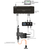
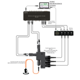
Bundle-imaging Fiber Photometry System
-
Conventional photodiode-based fiber photometry recordings of multiple brain sites on the same animal or single sites on several animals require a recording setup for each fiber location.
An elegant alternative to multiple site photodiode-based fiber photometry is CMOS- or sCMOS-based bundle-imaging fiber photometry. The setup resembles a fluorescence microscope with a fiber bundle end face in its focal plane. The individual fibers on the other end of the fiber bundle are fitted to the selected brain locations on a single animal or on different animals. The fluorescent light from each fiber within a bundle creates circular spots on CMOS. The electrical read-out from pixels within each of those spots or fiber images correlates with the calcium activity of the corresponding brain site.
For a Bundle Photometry option with optogenetics see: Bundle Photometry with Targeted Optogenetics (BFTO).
-
The GCaMP Bundle-imaging Fiber Photometry System records calcium-independent GCaMP fluorescence, excited by 405, 415, 425 or 440 nm light (isosbestic point), and its calcium-dependent fluorescence, excited by 470 nm light, from several fiber-connected brain sites with a CMOS image sensor. The sites labeled with GFP-like fluorophores can be on the same animal or distributed over many animals. The respective GCamP's excitations are interleaved or sequentially applied to separate calcium-dependent and calcium-independent fluorescence emissions.
The system includes:- 2-channel LED driver
- Bundle-imaging Fluorescence Mini Cube- GCaMP
-
Behavior & Bundle Photometry Console
- All required electrical cables
Optional items:- Fiber Photometry Cannula Holder to record during surgery
- Optical Power meter to monitor and calibrate excitation power
- Behavior Camera or CamLoop to record animal behavior
- Workstation
- danse™ analyzing software
- Pigtailed 1x1 Fiber-optic Rotary Joint with Holder (fixed or gimbal) for each animal in a separate cage with a single brain site
- Photometry Rack for BFPS
Accessories:- Low-autofluorescence Branching Fiber Patch cords to bring light to the cannula
- Fiber-optic Cannulas for chronic implant
- Mating sleeves for ferrule to ferrule interconnect
Note: Other light sources and fluorophore combinations are possible. Please enquire.
-
The GCaMP with Red Fluophore Bundle-imaging Fiber Photometry System records calcium-independent and calcium-dependent GCaMP fluorescence and Red Fluophore fluorescence from several fiber-connected brain sites with a single CMOS image sensor. Respective excitation wavelengths are 405, 415, 425 or 440 nm (isosbestic point), 470 nm, and 560 nm. The sites labeled with fluorophores can be on one animal or distributed over many animals. The respective fluorophore excitations are interleaved or sequentially applied to separate different fluorescence emissions.
The system includes:- 4-channel LED driver
-
Bundle-imaging Fluorescence Mini Cube- GCaMP with Red Fluorophore
-
Behavior & Bundle Photometry Console
- All required electrical cables
Optional items:- Fiber Photometry Cannula Holder to record during surgery
- Optical Power meter to monitor and calibrate excitation power
- Behavior Camera or CamLoop to record animal behavior
- Workstation
- danse™ analyzing software
- Pigtailed 1x1 Fiber-optic Rotary Joint with Holder (fixed or gimbal) for each animal in a separate cage with a single brain site
- Photometry Rack for BFPS
Accessories:- Low-autofluorescence Branching Fiber Patch cords to bring light to the animal
- Fiber-optic Cannulas for chronic implant
- Mating sleeves for ferrule to ferrule interconnect
Note: Other light sources and fluorophore combinations are possible. Please enquire.
-
-
Excitation wavelength 405, 415, 425 or 440 nm for GCaMP isosbestic excitation
470 nm for Green fluorophore excitation
560 nm for Red fluorophore excitationField of view 2.5 x 2.5 mm Maximum no. of sites 20x core 400 um NA0.37
60x core 200 um NA0.37
100x core 100 um NA0.37
* exact number of site could be limited by patchcord manufacturingExcitation Uniformity 10% over FOV Sensor High sensitivity CMOS image sensor (QE 86% @ 520 nm) Excitation Power Range typical 5 to 500 uW per fibre
(vary with optical fibre core and NA)Connectivity USB3.0 port on computer
SMA optical fiber bundle patch cords with adjustable focus (manual)Digital I/O 3 x Input or programmable outputs 5 V TTL Sampling Rate 3 to 20 Hz interleaved 3 excitations
up to 60 Hz for single wavelength excitationSoftware Doric Neuroscience Studio that synchronize with other Doric products as laser for optogenetics and behavior camera. -
Drawing Bundle-imaging Fluorescence Mini Cubes Rack- GCaMP
Bundle-imaging Fluorescence Mini Cubes Rack - GCaMP with RFP
User ManualFrEn Bundle-imaging Fluorescence Mini Cubes
- choosing a selection results in a full page refresh


