Fiber Photometry Solutions
Choose the right fiber photometry system for YOUR experimental needs.

All-in-one solutions
- Acquisition -> Data analysis
- Combine with behavior
- Compatible with optogenetics
- Plug-and-play
Fiber Photometry Systems
Fiber photometry is a neuroimaging technique that monitors neuronal activity in freely-moving animals. This technique utilises genetically encoded fluorescent indicators (e.g. GCaMP, dLight, GRAB-Ach, RCaMP, jRGECO1) expressed in target brain regions. These indicators fluorescence only when bound to melocules such as calcium, dopamine or acetylcholine, reporting real-time molecular dynamics during complex behaviors.
Unlike Doric Miniscopes, which resolve single-cell activity, fiber photometry records population signals (i.e bulk fluorescence from labelled neurons). But unlike microscopy, fiber photometry is minimally invasive, optimized for multi-animal & multi-site experiments, and more cost-effective.

Fiber photometry system are composes of several core components that, together, combine multiple wavelengths into a single or mutliple fiber(s) and collect the emitted fluorescence response, including:
- Console
- LED Driver & LEDs
- Fluorescence mini cube (FMC)
- Fiber-optic patch cords
- Rotary Joints (commutators)
- Fiber-optic cannulas
- Data Acquisition & Analysis Softwares
The latest generations of Doric systems integrate multiple components into a single device, providing several advantages. Learn more.
Browse below the role of each fiber photometry component:
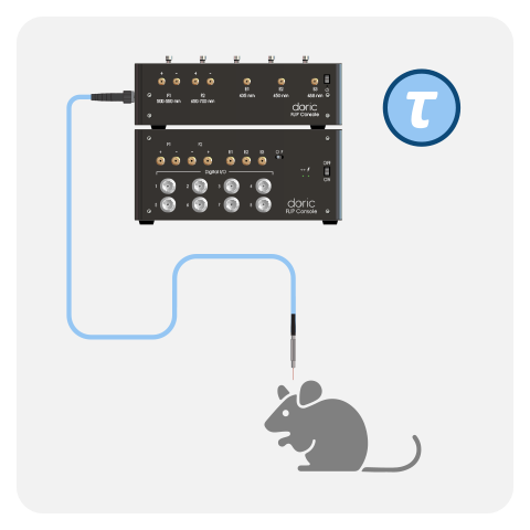
CONSOLE - data acquisition device
Consoles are data acquisition device that record the fiber photometry signal, control the timing and intensity of the LEDs and synchronize behavior, optogenetics, and other experimental components.
At Doric, we've designed several consoles over the years to address the needs of different fiber phometry systems. The Fiber Photometry Console (FPC), Behavior & Bundle Photometry Console (BBC300), and FluoPulse Console, are dedicated consoles for basic, bundle-imaging and fluorescence lifetime fiber photometry, respectively. Only the Neuroscience Console (NC500) is compatible mutltiple types of applications/fiber photometry systems.
All Doric console's come FREE with Doric Neuroscience Studio (DNS). Learn more.
FPC |
BBC300 |
NC500 |
FluoPulse™ |
||
|---|---|---|---|---|---|
| Max # site/animals | X4 X2 |
X 9-19* X 9-19* |
X8 X4 |
X2 X1 |
|
| Max # digital in/out | X4 | X32 | X32 | X4 | |
| FIBER PHOTOMETRY | Basic | ||||
| Rotary Basic | |||||
| Bundle-imaging | |||||
| Rotary Bundle | |||||
| Wireless | |||||
| Lifetime | |||||
| OPTOGENETICS | Close-Loop | ||||
| Wireless | |||||
| BEHAVIOR | Behavior Camera | ||||
| CamLoop** | |||||
| OTHER | Doric Miniscope | ||||
| Electrophysiology*** | |||||

1-color photometry + isosbestic (expect FluoPulse™ had no isosbestic)
2-color photometry + isosbestic (expect FluoPulse™ had no isosbestic)
* Depending on core fiber diameter: 9 x 400um, 19 x 200um
** Real-time animal tracking camera for close-loop experiments
*** Compatible with Intan RHD series
LEDS & DRIVERS - light source and its controler
In fiber photometry, LEDs are light sources that provide stable, wavelength-specific excitation (e.g., 405 nm, 465 nm, 560 nm, etc.) to selectively excite fluorescent indicators. An LED driver precisely controls the LED’s intensity and timing, enabling current-based modulation and synchronization with data acquisition for accurate demodulation/de-interleaving and separation of signals from different excitation wavelengths.
Both Basic and Bundle-imaging fiber photometry systems use low-power LEDs to prevent photobleaching and provide between 10-100 μW*.
In FluoPulse™ system, picosecond laser diodes (not LEDs) deliver an average power (1-30 μW).
In the latest generation of Doric fiber photometry systems, the light sources and drivers are directly integrated into the Fluorescence Mini Cubes.
*For 470nm with 400μm, 0.57NA patch cord.
Fluorescence Mini Cube - combine and seperate wavelengths
Fluorescence Mini Cube (FMC) are a critical fiber photometry component that combine the excitation from the light sources into a single fiber and seperates emission wavelengths towards the photodetector using dichroic mirrors and bandpass filters, preventing crosstalk between the wavelengths.
Over the years, the FMC design has integrated many core fiber photometry components including: the photodetectors & amplifier (iFMC), LEDs (ilFMC-G2) and LED Drivers (ilFMC-G3).
Compared to Fluorescence Mini Cubes without integration (FMC, Gen.1), photodetector integration (iFMC) provide a 100% better signal-to-noise ratio (30% signal increase and a 40% noise reduction). Moreover, ilFMC with integrated LEDs* and LED drivers reduce the number of connections and the system’s overall footprint, simplifying assembly and troubleshooting.
The ilFMC-G3 model, also has replaceable isosbestic LED (to adjust the isosbestic excitation for different biosensors without replacing the entire cube).
*The optogenetic light source is NOT integrated and must be included seperately.
NOTE: All Bundle-imaging and FluoPulse™ systems ONLY come in the all integrate format, such that mini cube also contains the photodetector, amplifier, light sources and drivers.

Rotary Joint / Commutator - optical joint for freely-moving animals
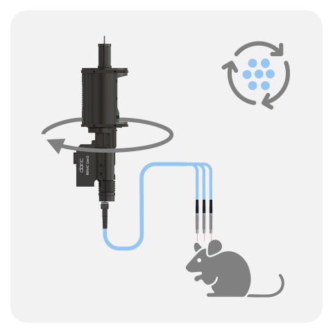
For freely-moving animals, we recommend rotary joints/optical communtators as they reduce damage to patch cords, tension on the animals head and prevent tangling. Single (FRJ_1x1_PT) or dual-fiber (rats: FRJ_2x2_PT or mice: AFRJ_2x2_PT) rotary joints can be added to most Doric fiber photometry systems and our ideal for long-term experiment lasting several hours or even days.
IMPORTANT: Pigtailed rotary joints (PT), with built-in fibers are required for fiber photometry to minimize rotation-related artifacts.
To completely ABOLISH rotation-related artifacts, we recommend rotary basic (1- or 2-sites) or Rotary Bundle Fiber Photometry Systems (3+ sites). In these systems, the detector (and fluorescence mini cube) is built-on to the rotary joint base, such that the detection happens during the rotation. Then it is the electrical signal from the detector that is transfered through the rotary joint (not the optical signal), completely by-passing the artifacts.
Fiber-optic Patch Cords - light delivery between system and animal
A fiber-optic patch cord is a flexible optical fiber assembly used to transmit light between system and the animal with minimal loss. Specifically, it delivers excitation light from the flight sources to the implanted optical fiber (cannula) and returns emitted fluorescence to the detector.
For fiber photometry applications, we recommend low-autofluorescence & flexible fiber-optic patch cords with high numerical apperture (NA). Silica/silica (0.37 NA) patch cords offer very low autofluorescence and high durability, providing cleaner signals and long-term stability but with lower light delivery and collection efficiency (i.e. lower NA). In contrast, Silica/polymer (0.57 NA) patch cords improve excitation and fluorescence collection due to higher NA, at the cost of reduced durability and moderately higher autofluorescence (must photobleach regularly).
For Bundle-imaging systems, we specifically recommend Silica/silica for its durability, to avoid replacing the more costly multi-fiber patch cords as often.
For the FluoPulse™ system, we strongly recommend Silica/Silica, as its very low autofluorescence provides the for best signal-to-noise ratio for fluorescence lifetime measures.
Fiber-optic Canulas - chronic fiber implant
A fiber-optic patch cord is a flexible optical fiber assembly used to transmit light between system and the animal with minimal loss. Specifically, it delivers excitation light from the flight sources to the implanted optical fiber (cannula) and returns emitted fluorescence to the detector.
For fiber photometry applications, we recommend low-autofluorescence & flexible fiber-optic patch cords with high numerical apperture (NA). Silica/silica (0.37 NA) patch cords offer very low autofluorescence and high durability, providing cleaner signals and long-term stability but with lower light delivery and collection efficiency (i.e. lower NA). In contrast, Silica/polymer (0.57 NA) patch cords improve excitation and fluorescence collection due to higher NA, at the cost of reduced durability and moderately higher autofluorescence (must photobleach regularly).
For Bundle-imaging systems, we specifically recommend Silica/silica for its durability, to avoid replacing the more costly multi-fiber patch cords as often.
For the FluoPulse™ system, we strongly recommend Silica/Silica, as its very low autofluorescence provides the for best signal-to-noise ratio for fluorescence lifetime measures.
Doric Lenses Inc. is a recognized leader in advanced fiber photometry solutions for behaving animals, driving innovation in this rapidly evolving field. Each system was carefully engineered to address the specific experimental needs & challenges.
The three main types of Fibers Photometry Systems are split into categories based on the type of detector.

Basic Systems
Classic photodetector system
Original technology.
Decade-long validation.

Bundle-imaging
One detector.
Multi-fibers.
Affordable multi-animal &
multi-site experiments.

Fluorescence Lifetime System
Picosecond changes
Quantify baseline & slow-state dynamics beyond intensity!
Explore how Doric’s fiber photometry systems compare across key metrics.
All Basic Fiber Photometry Systems are fitted with a high-sensitivity Doric photodetector. In the latest generations, the photodetector is integrated directly within the fluorescence mini cube / headstage (FMC, RFMC and WiFP), providing higher signal-to-noise ratio compared to non-integrated version. All Basic systems can be run in either lock-in mode (frequency-based division) or interleaved mode (time-based division) using live demodulation/deinterleave algorithms in (free) Doric Neuroscience Studio software.
Basic

- 1-2 sites/animal. Expand to 4-8 sites*
- Flexible Isosbetic point
- Simultaneous optogenetics
- Add rotary joint for freely-moving long-term behaviors
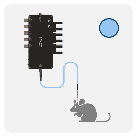
Rotary Basic

- 1-2 sites on ONE freely-moving animal
- Fixed isosbestic point
- Add optogenetic on non-photometry site
- Integrated rotary abolishes artifacts
Bundle-Imaging Fluorescence Mini Cube (BFMC-G3)
Ideal for long experiments (hours/days)
- Combine with fluid delivery
- Combine with electrophysiology / EEG

* Depending on the type of system and wether its 1-colour or 2-color configuration:
- Basic: 1-color (up to 8 sites/animals) and 2-colors (up to 4 sites/animals);
- Rotary: Max 4 animals (max 1-2 sites per animal, 1- or 2-colors);
- Wireless: Max 4 animals (1-site, 1-color per animal) .
One FMC/headstage can record the GFP-based and/or RFP-based signal from a single optical fiber. However, it is possible to combine multiple cubes/headstages to record from more than one mouse or brain site (up to 4-8*) when using the Neuroscience Console 500.
BASIC:
The latest generation of Basic Fluorescence Mini Cube (FMC) were disigned for flexible isosbestic point, such that the integrated isosbestic LED can be swapped between: 405, 415, 425 and 440 nm LEDs. In addition, both the 1-color and 2-color configuraitons are compatible with simultaneous red-shifted optogenetics, without crosstalk. The Basic system can be combined with pigtailed rotary joints (FRJ_1x1_PT, FRJ_2x2_PT & AFRJ_2x2_PT) for long-term, freely-moving experiments. With decade-long validation, the basic fiber photometry system is ideal to record GFP, RFP and isosbestic signals from 1-2 sites/animals, with the possibility to expand later on.
ROTARY BASIC:
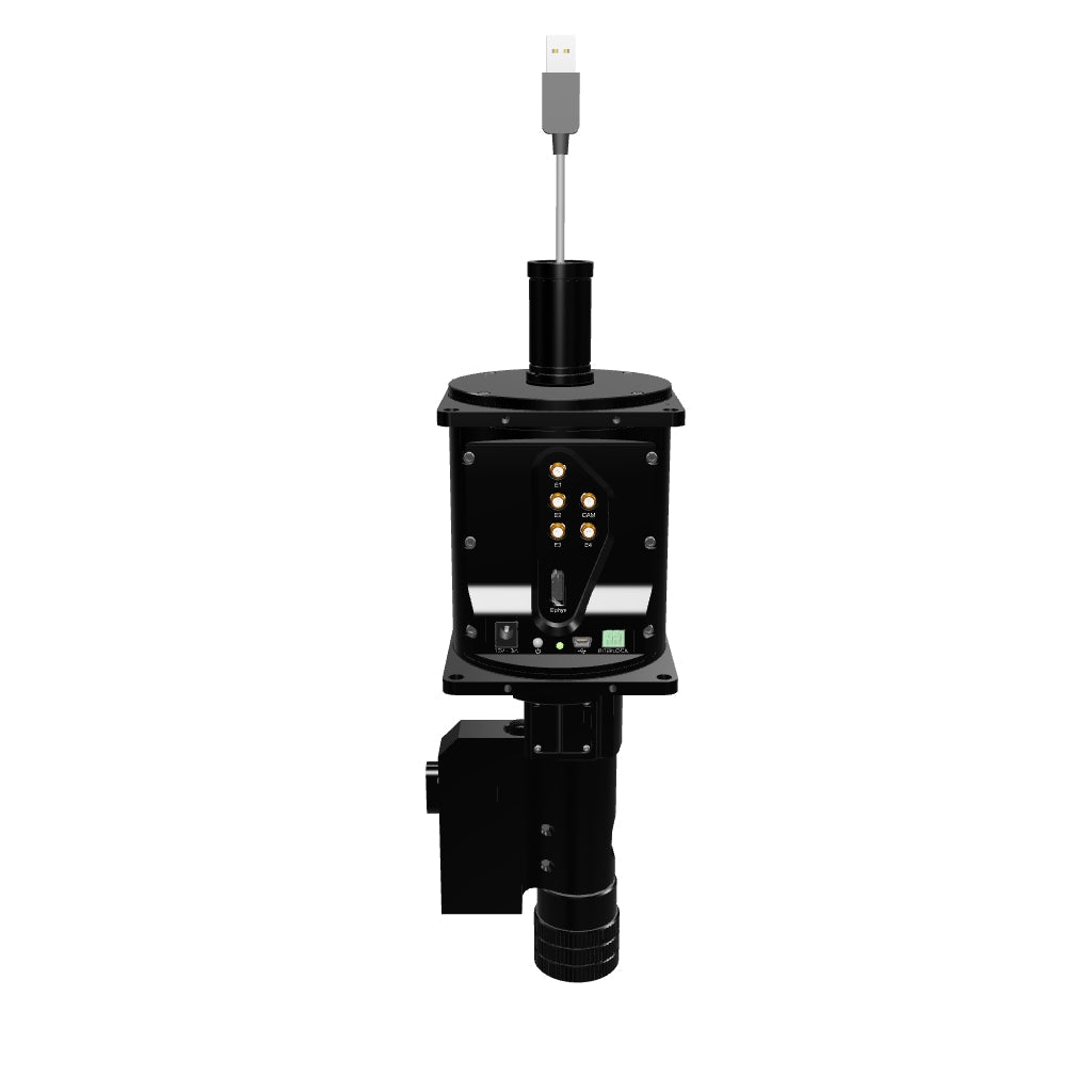
In Rotary Fiber Photometry Mini-Cube (RFMC), up to two fluorescence mini cube(s) are built-on the rotary joints itself. This unique design abolishes rotational artifacts in the signal and minimizes the number of connections before the photodetector. Therefore, RFMC system boasts a higher quality signal compared to basic FMC system paired with an external rotary joint, and is ideal for experiments lasting several hours or even days. The rotary joint also contains a hollow channel that can be used for fluid delivery, electrophysiology/EEG, or optogenetics.
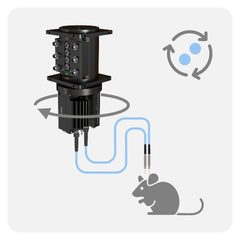
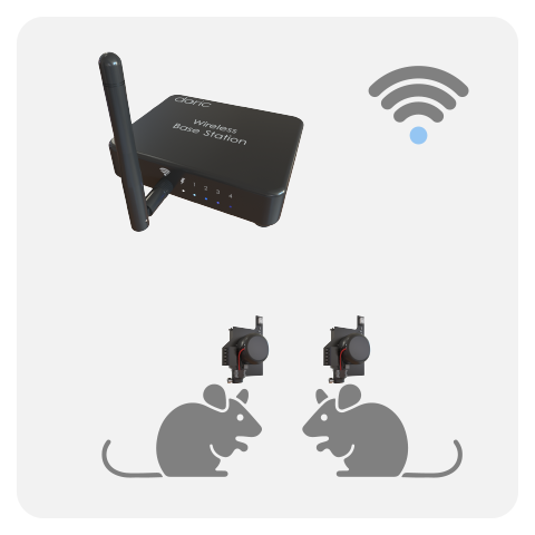
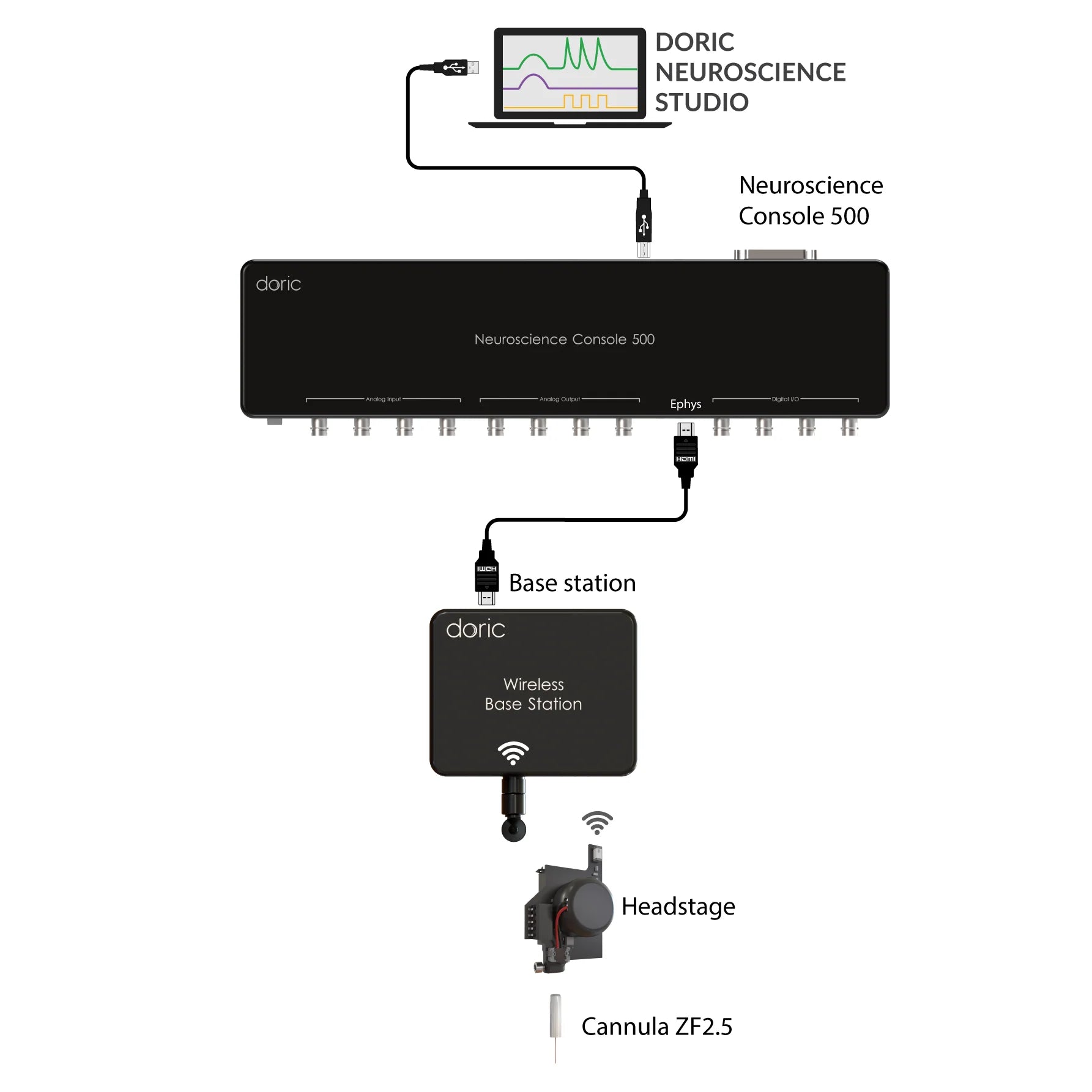
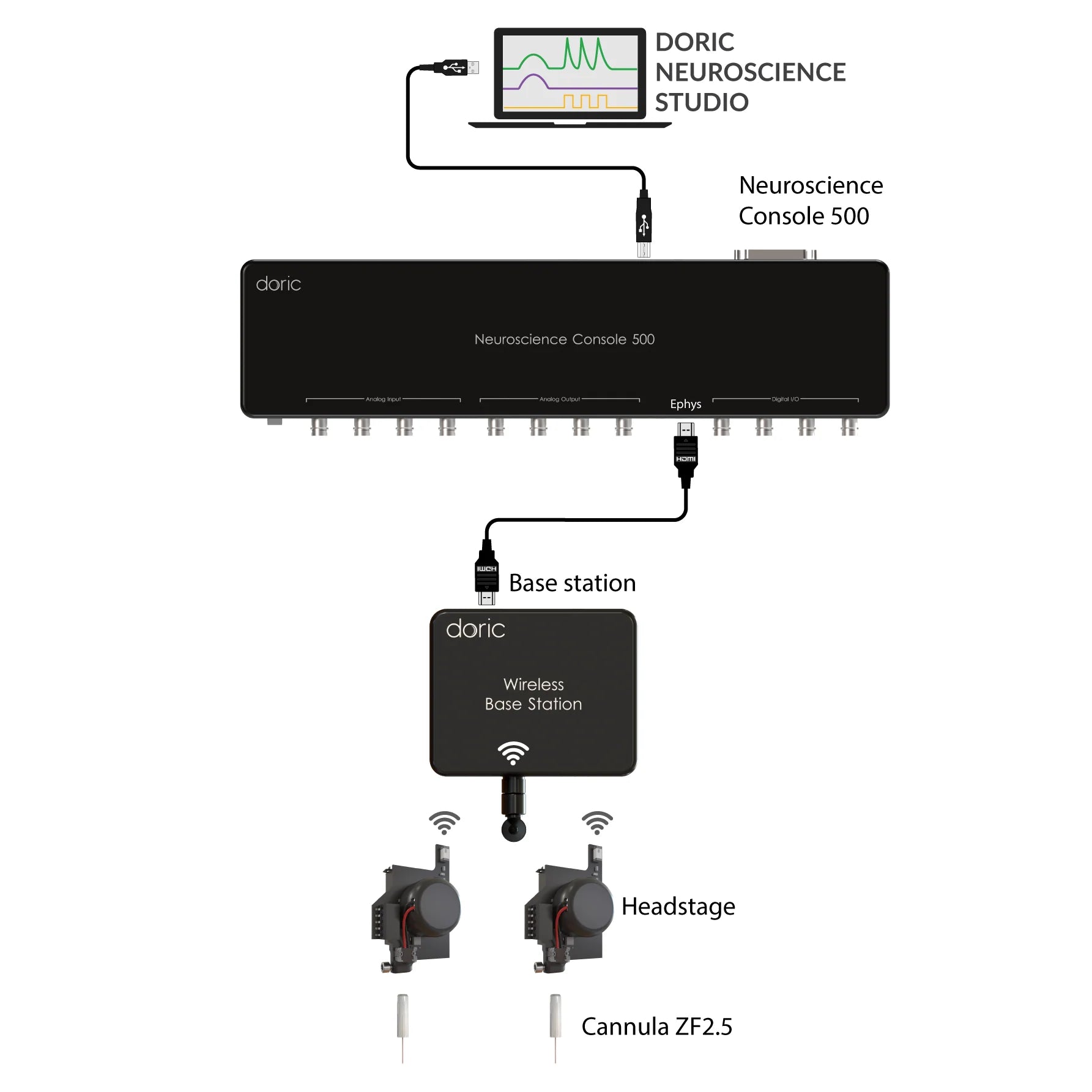
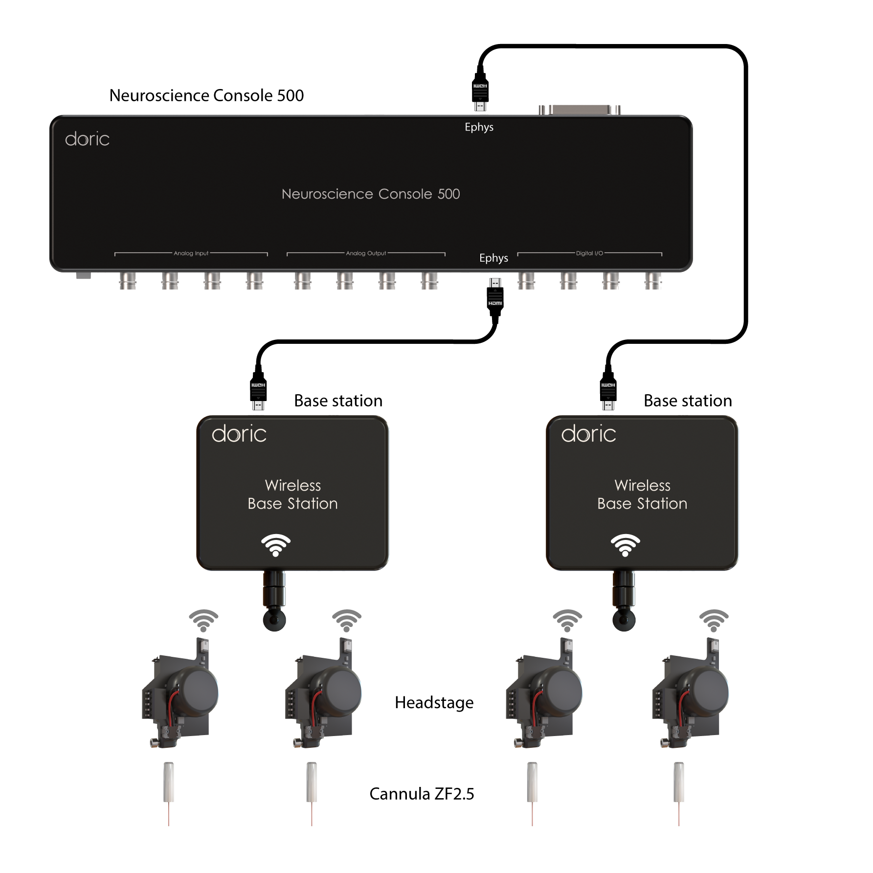
WIRELESS:
Lastly, the Wireless Fiber Photometry System (WiFP) was designed to circumvent tangling issues when recording multiple animals in the same cage or for behaviors disrupted by tethered system. The ilFMC4 (with 470 and isosbestic LED and detector) were miniturized onto a lightweight headstage. Multiple headstages can be run at the same time, thus this system is ideal to study neural population underpinning social behaviors.
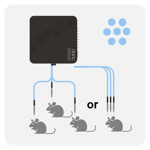
Unlike the Basic Fiber Photometry Systems, which require one mini cube per site/animal, the Bundle-imaging Fiber Photometry systems use a CMOS camera to image the entire fiber bundle simultaneously. Bundle-Imaging Fiber Photometry System are ideal when recording 1- or 2-colors from multiple animals or brain regions (1-19 fibers), all at a reasonable price.
All bundle-imaging system are available with 2-color configurations (plus isosbestic) and the three excitations are interleaved in time (i.e the lock-in mode is not available). All three LEDs excite the entire fiber bundle (no independent power adjustement), and the photometry signal is then sampled at 20-100 Hz depending on the field-of-view of the camera and the number of fibers imaged at the same time.
Three bundle-imaging system options differ based on the level of integration and compatibility with optogenetics:
Bundle-imaging
Bundle-imaging
Fluorescence Mini Cube (BFMC)

- Multi-animal
- Multi-site
- 2-color fiber photometry
- Fixed isosbestic point
- No simultaneous optogenetics
- Add external rotary joint for freely-moving behviors
- Plug-and-play
Bundle-imaging + Opto
Bundle-imaging with
Targeted Optogenetics (BFTO)

- Multi-animal
- Multi-site
- 2-colors fiber photometry
- Fixed isosbestic point
- Independent, multi-site optogenetics
- Add external rotary joint for freely-moving behviors
Rotary Bundle-imaging
Rotary Bundle-imaging
Fluorescence Mini Cube (RBFMC)

- Single animal
- Multi-site
- 2-colors fiber photometry*
- Flexible isosbestic point
- Compatible with simultaneous optogenetics*
- Integrated rotary abolishes artifacts
- Ideal for long experiments (hours/days)
- Compatible with electrophysiology / EEG
BUNDLE-IMAGING (GEN.3):
The BFMC-G3 is fully integrated, plug & play design (with CMOS camera, LEDs, LED Driver & console in a single device), greatly simplifying the set-up & troubleshooting. However, this system is not compatible with simultaneous optogenetics.
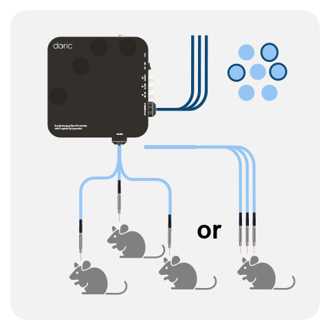
BUNDLE-IMAGING WITH TARGETED OPTOGENETICS (BFTO):
The BFTO system is specifically designed for multi-fiber photometry with targeted optogenetics. The term "targeted" refers to independent, multi-site optogenetic control is ideal for close-loop optogenetics (both multi-animal and/or multi-site) experiments. This system provides the greatest flexibility for common neuroscience experiments.
ROTARY BUNDLE-IMAGING:
The RBFMC is designed for single-animal, multi-site experiment lasting several hours/days. The CMOS detector is integrated directly on the rotating joint ABOLISHING rotation-related artifacts, allowing for high-quality signals. The 2025 version now comes with a flexible isosbestic design, easily switching between different biosensors. Additionally, the RBFMC includes optogenetic capabilities that can illuminate all sites simultaneously* or can be fitting with external laser diode for independent optogenetic stimulation on a non-fiber photometry site.
* Simultaneous optogenetic on the fiber photomery sites is only available for the 1-color configuration; For 2-color configuration optogenetics can be added, but on a non-photometry site
The latest innovation in fiber photometry!
Fluorescence lifetime fiber photometry measures changes in the fluorescence decay time of indicators rather than signal intensity, providing a readout that is largely independent of motion artifacts, photobleaching, and fiber coupling fluctuations. This makes it especially powerful for stable, tonic or baseline changes in neural activity or biochemical states in freely-behaving animals.
FluoPulse™
Fluorescence lifetime Fiber Photoemtry Cube (FLPC)

- Measure Fluorescence Lifetime (ns)
- 1-color or 2-color*
- 1-2 animals
- Simultaneous optogenetics same site
- Robust to motion artifacts
Ideal for long experiments (hours/days) - Measure tonic/baseline line changes
WHAT IS FLUORESCENCE LIFETIME?
Fluorescence lifetime is the average time a fluorophore remains in its excited state before emitting a photon, typically on the nanosecond timescale. It is an intrinsic property of the fluorophore that depends on its local molecular environment rather than its concentration or excitation intensity.

Want to learn more?
Register for the Discover FluoPulse webinar, run quartely (or request a recording).
SYSTEM COMPARISON
This side-by-side comparison highlights differences in channel configuration, maximum number of site/animal(s), dual-color & optogenetics compatibility, and advanced features across seven distinct fiber photometry systems.
| BASIC | BUNDLE-IMAGING | LIFETIME | ||||||
|---|---|---|---|---|---|---|---|---|
|
|
|
|
|
|
|
|
||
| Fiber Photometry |
Flexible isosbestic | ✔ | ✔ | n/a | ||||
|
Max # sites
●
◐
|
Up to 8 Up to 4 |
1–2 | 1 ✖ |
Up to 9–19* | Up to 9–19* | Up to 7–19* | 1–2 1 |
|
|
Multi-animal (separate cage)
●
◐
|
Up to 8 Up to 4 |
Up to 4 ✖ |
Up to 9–19* | Up to 9–19* | 1–2 ✖ |
|||
|
Multi-animal (same cage)
●
|
Up to 4 | |||||||
| Optogenetics | Same site as photometry | ✔ | ✔ | ✔*** | ✔ | |||
| Different site as photometry | ✔ | ✔ | ✔ | ✔ | ✔ | ✔ | ||
| Behavior (Rotary joint / commutator) |
Long-term, freely-moving behavior | ** | ✔ | 2–4 h | ** | ** | ✔ | ✔ |
| Integrated rotary joint | ✔ | n/a | ✔ | n/a | ||||
| Interacting animals | ✔ | |||||||
| Add Ephys / EEG & EMG | ** | ✔ | ** | ** | ✔ | ** | ||