Doric Miniscope Solutions
All-In-One Solution
What is miniscope?
Fluorescence Miniscope system images single-cell activity, in freely-behaving animals. Unlike two-photon and confocal microscopy, which requires head-fixation or anesthesia, miniaturized microscopy enables studying unrestrained behavior by directly attaching a small, lightweight microscope to the animal’s head. This technique utilizes genetically encoded fluorophore indicators, such as GCaMP, dLight, and GRAB-Ach, expressed in the target brain region. These indicators fluoresce when they bind specific molecules (e.g., dopamine, acetylcholine, calcium ions), serving as a proxy for neuronal activity. Thus, the miniaturized microscope is the ideal method to study the neuronal correlates of complex behaviors, in real time.

Since 2014, Doric Lenses has been among the leaders in the development of fluorescence miniscope systems and provides a comprehensive, all-in-one solution: from the implant to data analysis. This includes imaging cannulas, microscope body, rotary joint (commutator), Fluorescence Microscope Driver, and data analysis solution (danse™). The Doric miniscope systems are compatible with optogenetics and behavior acquisition (Behavior Camera/CamLoop), ensuring a seamless workflow.
As of today, several types of microscope bodies are carefully developed at the company to provide the best imaging quality based on the depth of the targeted brain structure. In this regard, Doric miniscopes can generally be categorized into two distinct sets:
• Surface Miniscope – Specifically designed for imaging superficial brain structures including all cortical areas.
• Deep Brain Miniscope – Optimized for all depth imaging including the deep brain structures, with the flexibility to also be used for surface area imaging.
See image below, which categorizes all Doric fluorescence miniscope systems:

Miniscope Options
Surface Miniscope
The Surface Miniscope is designed for imaging superficial brain regions located up to 150 mm deep, such as the Visual Cortex and Prefrontal Cortex. These microscopes are combined with a cranial window above the region of interest, which then connects to the microscope body via snap-in connection. This cranial window design offers two key advantages:
1- Reduced Surgery invasiveness – A significantly easier and less invasive procedure, addressing one of the most challenging steps in miniature microscopy.
2- Superior Signal Quality – Larger diameter optical components provide higher image resolution across the entire field of view (FOV).
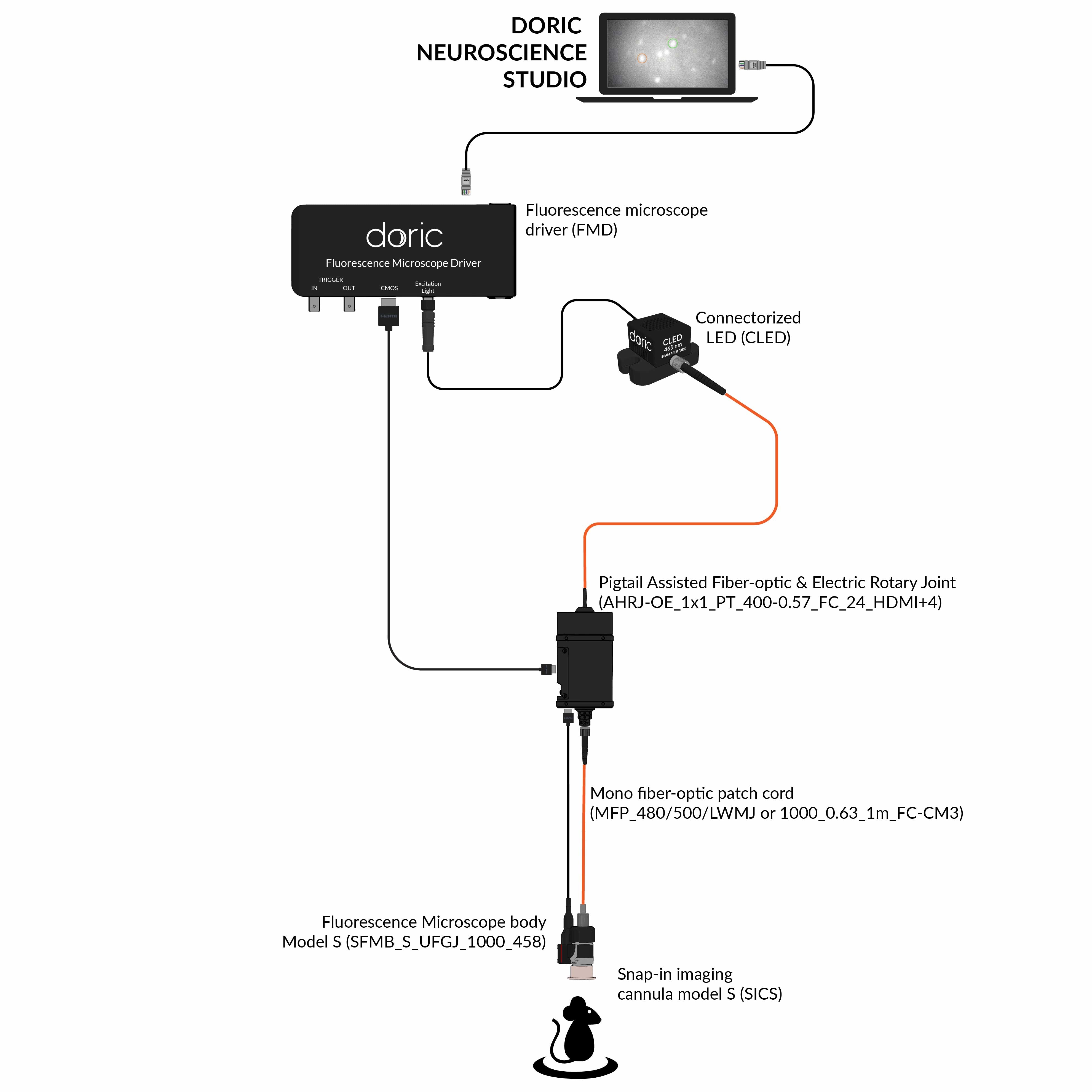
Surface Miniscope set includes the following versions:
[Snap-in (SFMB-S)] → 1-color imaging (green or red fluorescence)
[OSFM-S] → 1-color (green fluorescence) imaging with optogenetics (yellow-red wavelength) OR 1-color (red fluorescence) imaging with optogenetics (blue wavelength)
[2CFM-S] → 2-color imaging (green and red fluorescence)
The 1-color + optogenetics microscope (OSFM-S) is capable of providing up to 55 mW / mm2 of yellow-red wavelength light (580-640 nm pulsed or continuous) at the tip of the implant. This high-power output ensures efficient activation of yellow-red activated opsins, such as Halorhodopsin, which can be challenging to stimulate effectively.
Despite advantages, the larger optical lenses used in surface Miniscope systems are not suitable for deep brain imaging, as they may cause significant tissue damage if intruded deeply in brain. Even though specific custom configurations are proposed for imaging deeper layers of cortex and hippocampus using Surface Miniscope, usually a dedicated solution for deep structures imaging is required.
Snap-in SFMB-S

OSFM-S

2CFM-S

Deep Brain Miniscope
Deep brain Miniscope solution is designed to record neural activity from structures located deep within the brain that cannot be accessed using the SURFACE Miniscope. These include regions such as the Substantia Nigra, Hypothalamus, and Parabrachial Nucleus, among many others. Deep brain Miniscope has a distinct optical design which requires GRIN lens (thin rod-like lens) implantation for imaging purposes. To simplify the surgery and ensure an efficient optical alignment, the GRIN lens is integrated into an imaging cannula. This microscope set includes the following versions:
The first two Deep brain Miniscopes, 1-color (eTFMB3) and 1-color+optogenetics (eTOSFM3), are also known as Twist_on microscopes. This name comes from the connection mechanism to the cannulas (no pressure on the skull during connection, no tools required). They are also referred to as eFocus in the literature because they contain an embedded electronically adjustable lens inside the microscope body. Such electronic focus can be adjusted in the Doric Neuroscience Studio (DNS) software with a depth movement range of 300 µm, enabling remote focus adjustments between sessions and ensuring optimal imaging quality. The efocus feature provides a way to track the same cells over time and improves fluorescence signal and spatial resolution.
The 1-color + optogenetics microscope (eTOSFM3) provides up to 55 mW / mm2 of yellow-red wavelength light (580-640 nm pulsed or continuous) at the tip of the implant. This high-power output ensures efficient activation of yellow-red activated opsins, such as Halorhodopsin that have higher activation thresholds.
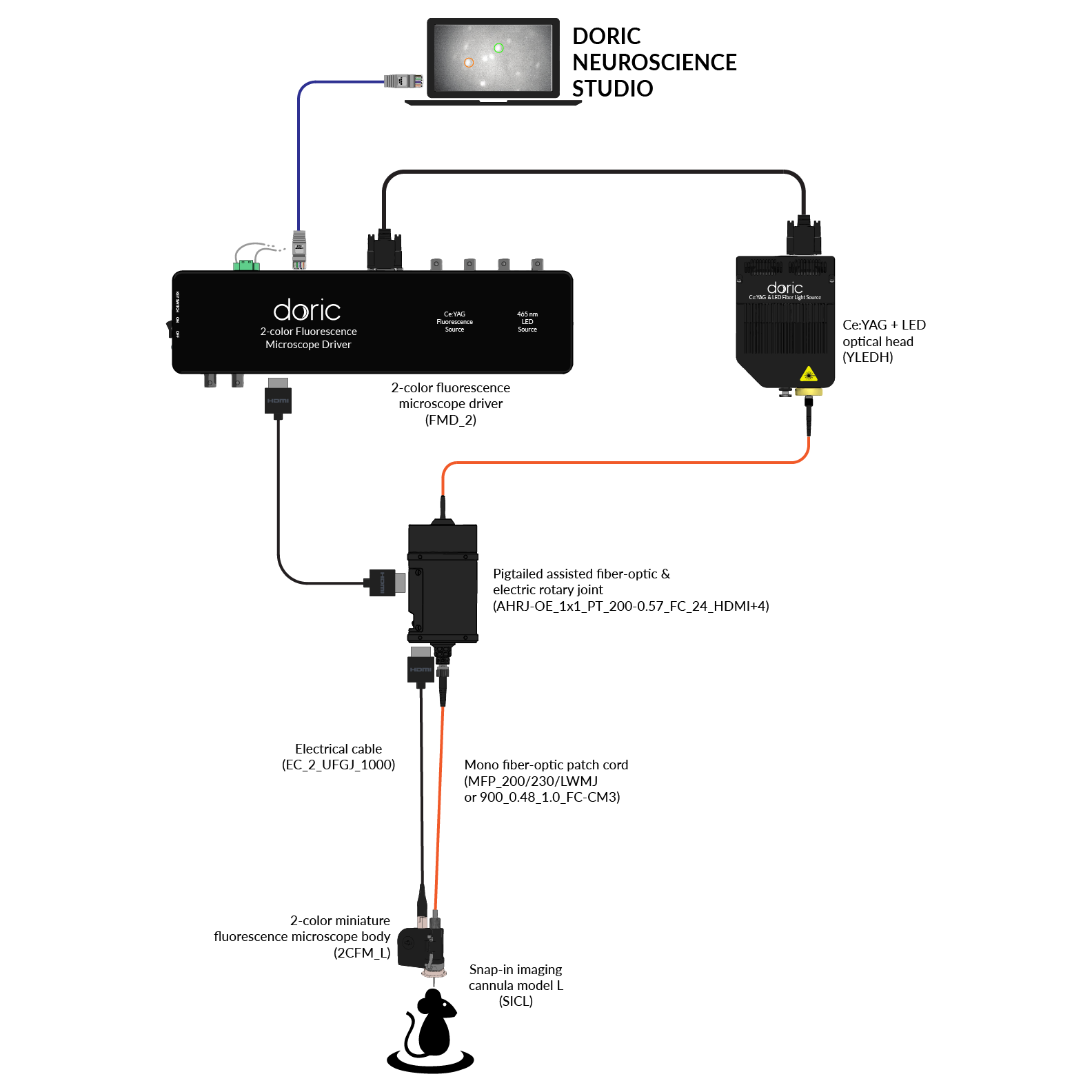
Lastly, the 2-color microscope (2CFM-L), features two separate integrated CMOS sensors, each dedicated to green and red color imaging. To ensure that both colors are imaged in the same plane, users need to specify the brain region of interest. The LD version supports 2-color imaging at 0-3.4 mm in depth, and LV version supports 2.9-5.9 mm depths.
eTFMB3
eTOSFM3
2CFM-L
Explore Additional Modalities
Using Rotary Joint (Commutator)
Miniscopes are typically wired, and during animal movement the tether can twist and become tangled. This tangling can significantly restrict the animal’s mobility and potentially affect behavioral performance. A common solution is to use a rotary joint (commutator), which compensates for tether twisting and helps relieve tension and pressure on the animal during free movement experiments.
For Doric miniscope imaging, the recommended compatible rotary joint is the Assisted 1*1 Pigtailed Fiber-optic & Electric Rotary Joint – 24 contacts. This rotary joint has:
- An optical path for delivering excitation light required for miniscope imaging
- An electrical path for transmitting the recorded neural imaging signal to the acquisition system
Note that the rotary joint is available with different electrical connector types. For full compatibility with Doric miniscopes, the HDMI connector version should be selected.
A major advantage of this rotary joint is its versatility. The optical channel can also be used for optogenetics or fiber photometry recordings in other experiments, while the electrical channel can support transmission of ephys signals. In other words, it is a multi-purpose investment; one rotary joint can support multiple experimental workflows.
This rotary joint is also available in a 2×2 version, which includes two separate optical paths. While one optical path would be used for miniscope imaging, the second path could be used for optogenetics at a different brain site (or other applications).
Additionally, these rotary joints are relatively compact (approximately 8.5 cm in height) and operate with low acoustic noise, helping reduce disruption during sensitive behavioral and neuroscience experiments.
Combining real time-animal tracking and closed-loop optogenetic
We recommend using the Doric CamLoop camera for real-time animal tracking and closed-loop optogenetic stimulation.
To enable this functionality, the camera must be paried with with the FMD3 or LaserTM/FMD, or NC500 drivers. For compatibility with these consoles, Deep Brain Miniscope 1-color or 1-color + optogenetic models should be used.


Lateral imaging with prism
Typical miniscopes image cell populations located directly beneath their flat imaging windows (cranial widow or GRIN lens depending on the system). However, upon custom-request, all Doric microscopes can be coupled with a prism mirror to enable lateral imaging.
This configuration offers two key advantages:
- Layered Imaging – Collect data from multiple layers simultaneously from a brain structure, providing a more comprehensive view of neural activity.
- Reduced Tissue Damage – The sharp edge of the prism glass minimizes tissue damage and immune response after implantation, compared to traditional blunt probes.
This prism-assisted imaging method has been successfully implemented by customers for imaging both deep and superficial brain structures. SEE PUBLICATIONS.

Fluid Injection
For certain experiments, researchers may need to perform fluid injections at the same location as microscopy imaging or at a different brain site. All Doric Deep Brain Miniscopes are compatible with in-house produced opto-fluidic cannulas, specifically designed for these experiments.
**Note: In these setups, the standard rotary joint may no longer be compatible with the microscope. To find alternative solutions, please contact us.

Combining other methods with miniscope imaging
When combining a miniscope with other optic manipulations is needed, we highly recommend using our latest acquisition console, Neuroscience Console 500 (NC500). This advanced system supports real-time data collection from multiple modalities, including:
- Doric Efocus Miniscope
- Fiber Photometry Systems (Basic, Bundle, Wireless)
- Optogenetics
- Behavior Camera
- CamLoop Camera for real-time tracking in closed-loop optogenetics
- Electrophysiology (Intan RDH series)

User-friendly data acquisition software

Doric Neuroscience Studio
FREE data acquisition software
Intuitive software with multiple modalities for controling all doric devices, from minisope to photometry, behavior and optogenetics.
All-in-one data analysis software
danse™ - data analysis solution
Automate Find Cell with integrated CaImAn, MiniAn, or Suit2p and streamline your parameter selection with multiple preview steps, without any coding required!Align cell traces with animal behavior, for batch processing all in one software.
