
VoluScan™
VoluScan™
VoluScan™ is a fluorescent mesoscope that enables 1- or 2-color functional imaging of large sample volumes at a cellular resolution. VoluScan™ is designed to image fluorescent sensors (like GCaMP, jRGECO, dLight, etc.) in organoids, assembloids, brain slices, and cell cultures, but can also be used to study cell migration, division, or other processes. Furthermore, the system is compatible with Tokai Hit incubation chamber and can synchronize imaging with other systems using external digital triggers. Thus, VoluScan™ pairs readily with Light Sources (for optogenetics or uncaging), electrophysiology recordings, liquid infusion, etc.
VoluScan™ has an extensive field of view (up to 5 mm) with subcellular resolution (2 µm), and covers a large depth (up to 1.5 mm). Specifically, the VoluScan™'s multi-plane imaging system enables scanning such volumes at high sampling rates.
…generating
VoluScan™ is a fluorescent mesoscope that enables 1- or 2-color functional imaging of large sample volumes at a cellular resolution. VoluScan™ is designed to image fluorescent sensors (like GCaMP, jRGECO, dLight, etc.) in organoids, assembloids, brain slices, and cell cultures, but can also be used to study cell migration, division, or other processes. Furthermore, the system is compatible with Tokai Hit incubation chamber and can synchronize imaging with other systems using external digital triggers. Thus, VoluScan™ pairs readily with Light Sources (for optogenetics or uncaging), electrophysiology recordings, liquid infusion, etc.
The VoluScan™ has an extensive field of view (up to 5 mm) with subcellular resolution (2 µm), and covers a large depth (up to 1.5 mm). Specifically, the VoluScan™'s multi-plane imaging system enables scanning such volumes at high sampling rates:
- - 4-planes (1-color) at 16 FPS
- - 8-planes (1-color) at 10 FPS
Each VoluScan system includes a VoluScan's multi-plane disc that determines the number of imaging planes (4 or 8), the z-stack steps, and the total imaging depth. By default, the z-stack increments are available in 50 µm, 100 µm or 150 µm steps (others on custom request). These discs are interchangeable to fit the needs of different experiments and can be purchased separately.
Check out the first bioRiv publication of assembloid data collected with VoluScan™. Miura et al. (2024), used the VoluScan's large FOV and fast z-scanning capabilities to image the live, calcium dynamics of over 400 cells per assembloid and demonstrated a functioning model of the cortico-striatal-thamalic-cortical circuit. Learn more
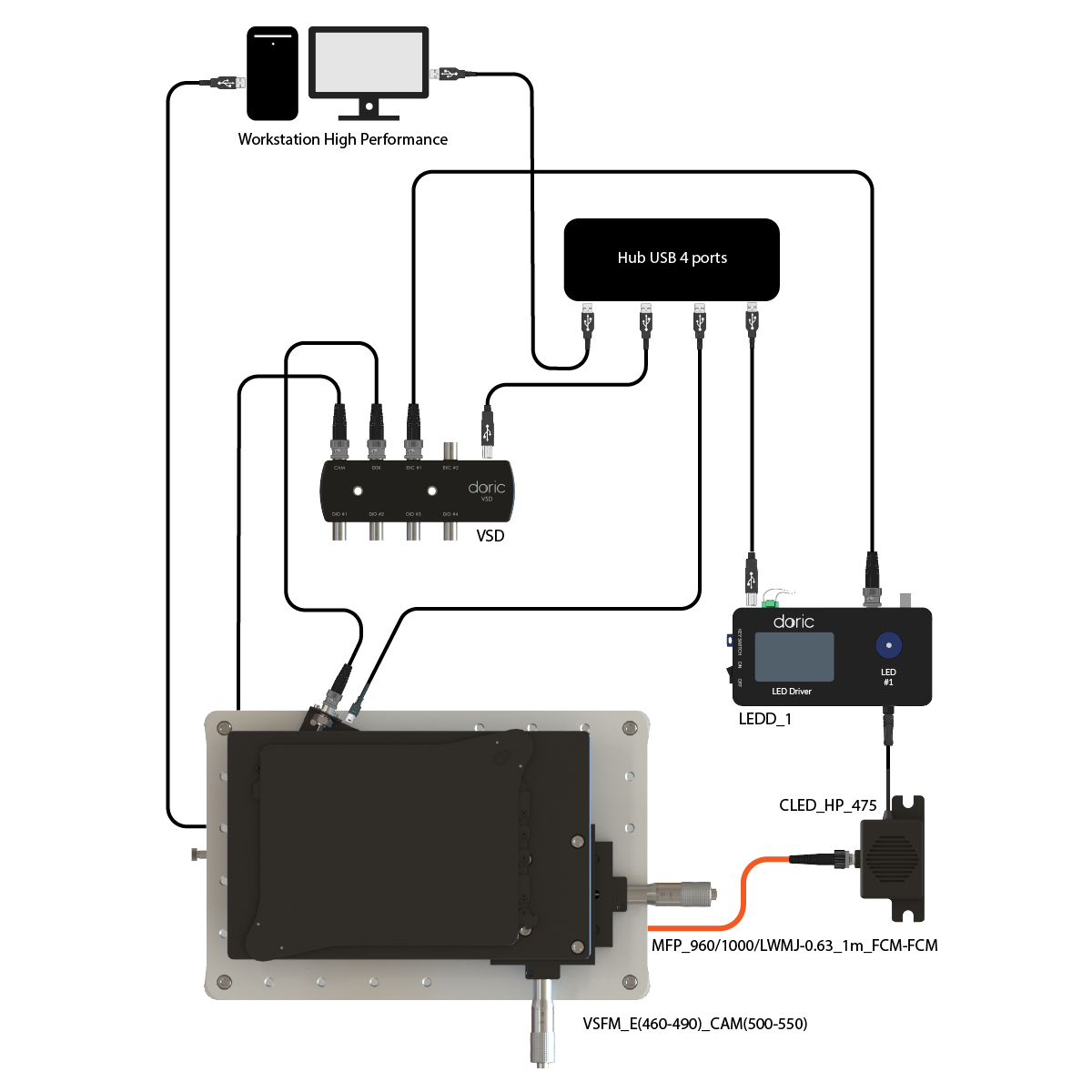
The VoluScan™ system for GCaMP imaging includes:
- Voluscan™ Gen1
- Voluscan™ multiplan disk
- VoluScan™ Driver
- LED Driver 1 channel to control LED power
- Connectorized LEDs - High Power at 470 nm
- Workstation - high performance
- All necessary cables
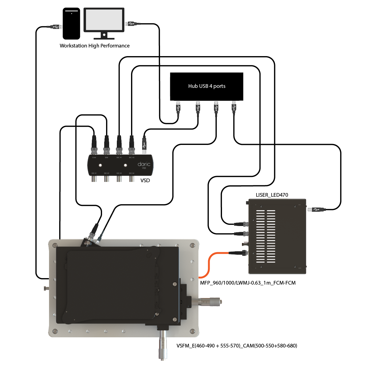
The VoluScan™ system for GCaMP imaging includes:
- Voluscan™ Gen1
- Voluscan™ multiplan disk
- VoluScan™ Driver
- Liser™light source with LED 475
- Workstation - high performance
- All necessary cables
| General properties: | |
|---|---|
| Field of view | 3.2 mm x 5.0 mm |
| Objective NA | 0.16 |
| Magnification | 2.2x |
| Working distance | 5 mm |
| Configuration | inverted* |
| Mechanicals stage travel | X: 25 mm Y: 25 mm Z: 12.5 mm |
| Dimensions | 210 x 320 x 400 mm |
| Computer interface | 1x USB2.0 port + 1x USB3.0 port |
| Z stack properties | |
| Number ofplanes | 4, 8 |
| Step | 50, 100 or 150 µm** |
| Maximum depth range | 1.5 mm |
| Sensor | |
| Type | CMOS Image Sensor*** |
| Quantum efficiency | 82% at 520 nm |
| Resolution | 1920 x 1200 pixels |
| Pixel Size | 5.86 µm x 5.86 µm |
| 1-color | |
| Spectral bandwidth | Excitation: 460-490 nm Emission: 500-550 nm |
| Maximum framerate (full FOV) | 1 plane: 80 FPS 4 planes: 16 FPS 8 planes: 10 FPS |
| 2-color | |
| Spectral bandwidth | Excitation #1: 400-410 nm or 410-420 nm Excitation #2: 555-570 nm Emission #1: 500-540 nm Emission #2: 580-680 nm |
| Maximum framerate (full FOV) | 1 plane: 40 FPS 4 planes: 8 FPS 8 planes: 5 FPS |
| DI/O | |
| Number of ports | 4 |
| Input voltage | 0 to 5.0 V (Maximum) |
| TTL level | HI > 3.3 V LOW < 1.5 V |
| Output voltage | TTL 5.0 V (High impedance) |
| Output Current | max 20 mA per channel |
| Maximum sampling rate | 10 kSps |
| Maximum output frequency | 5 kHz |
** other value on custom request
***Andor sCMOS on custom request
Related products
-
Choose your option
