Snap-in Surface Fluorescence Microscopy systems
Snap-in Surface Fluorescence Microscopy systems
Surface snap-in microscopy systems are optimized to image brain structures up to 150 um deep without using invasive implants. As no GRIN lenses are being used, the image quality of surface microscopes is higher.
Surface snap-in microscopy systems are optimized to image brain structures up to 150 um deep without using invasive implants. As no GRIN lenses are being used, the image quality of surface microscopes is higher.
Motor assisted rotary joint (AHRJ) and microscope dummy are available on option.
The GCaMP surface microscopy system is optimized for GFP-like fluorescence proteins imaging.
The system includes:

Add optional items:
Complete your system with:
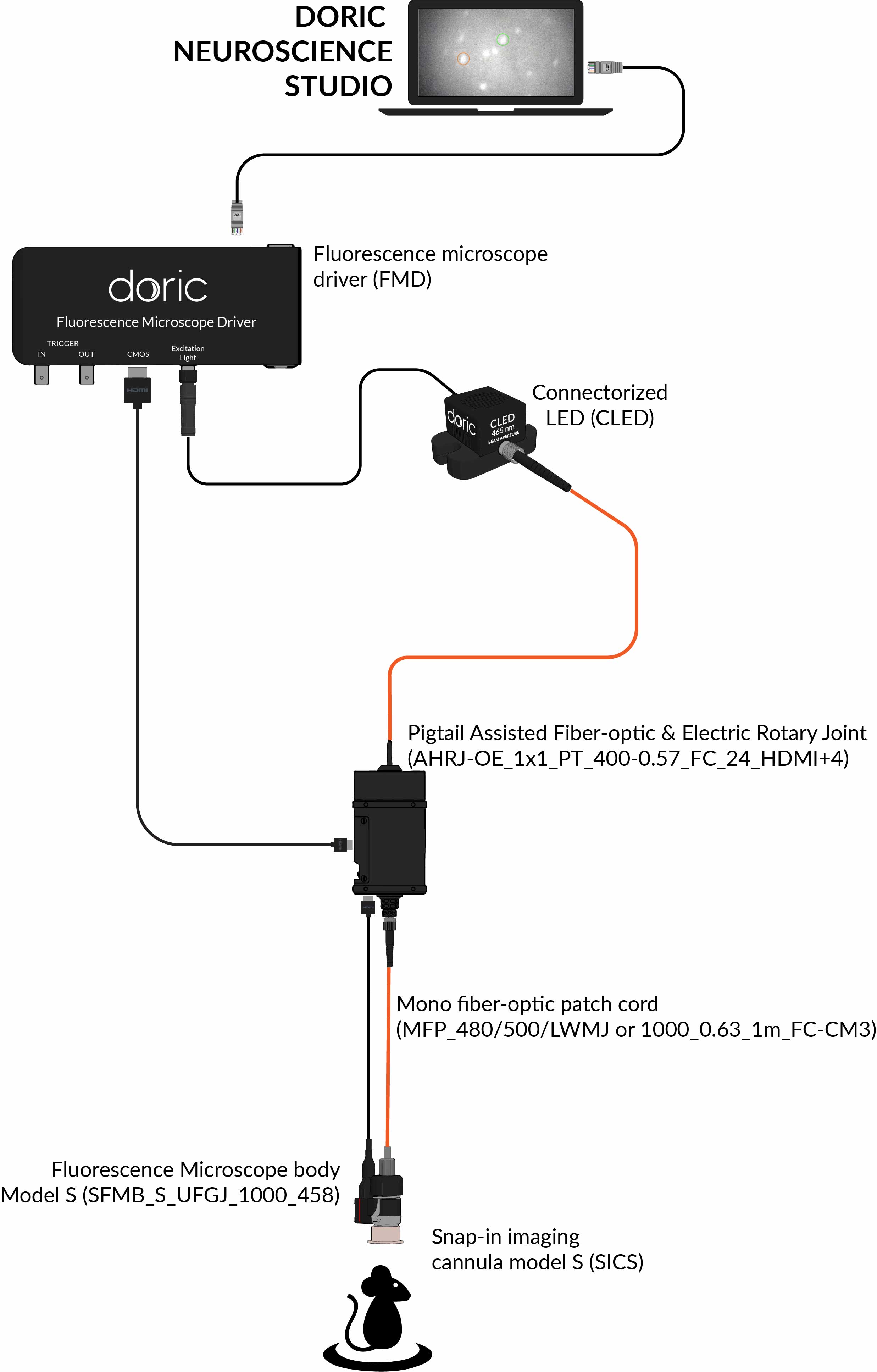
The system includes:

- Snap-in Green Fluorescence Microscope Body, Model S, the head mounted microscope
- Fluorescence Microscope Driver, to control the microscope
- Connectorized LED
- Optical fiber patch cords, to connect the light source to the microscope
- Fluorescence Microscope Holder
- Clamp for Fluorescence Microscope Holder
- Doric Neuroscience Studio Software
Add optional items:
- Assisted 1x1 Pigtailed Fiber-optic & Electric Rotary Joint - 24 contacts
- Snap-in Microscope Dummy, Model S
- Behavior Camera to record animal behavior
- Workstation
- danse™ analyzing software
Complete your system with:
| Field of View | 700 µm x 700 µm |
| Working distance | 1.1 mm |
| Frame rate | > 45 fps |
| Fluorescence recording | |
|---|---|
| Excitation | 458 / 35 nm |
| Emission | 525 / 40 nm |
The GCaMP + Red Opsins Surface Microscopy System is optimized for GFP-like fluorescence proteins imaging and Red Opsins (e.g. NpHR3.0, Chrimson, ...) activation.
The system includes:

Add optional items:
Complete your system with:
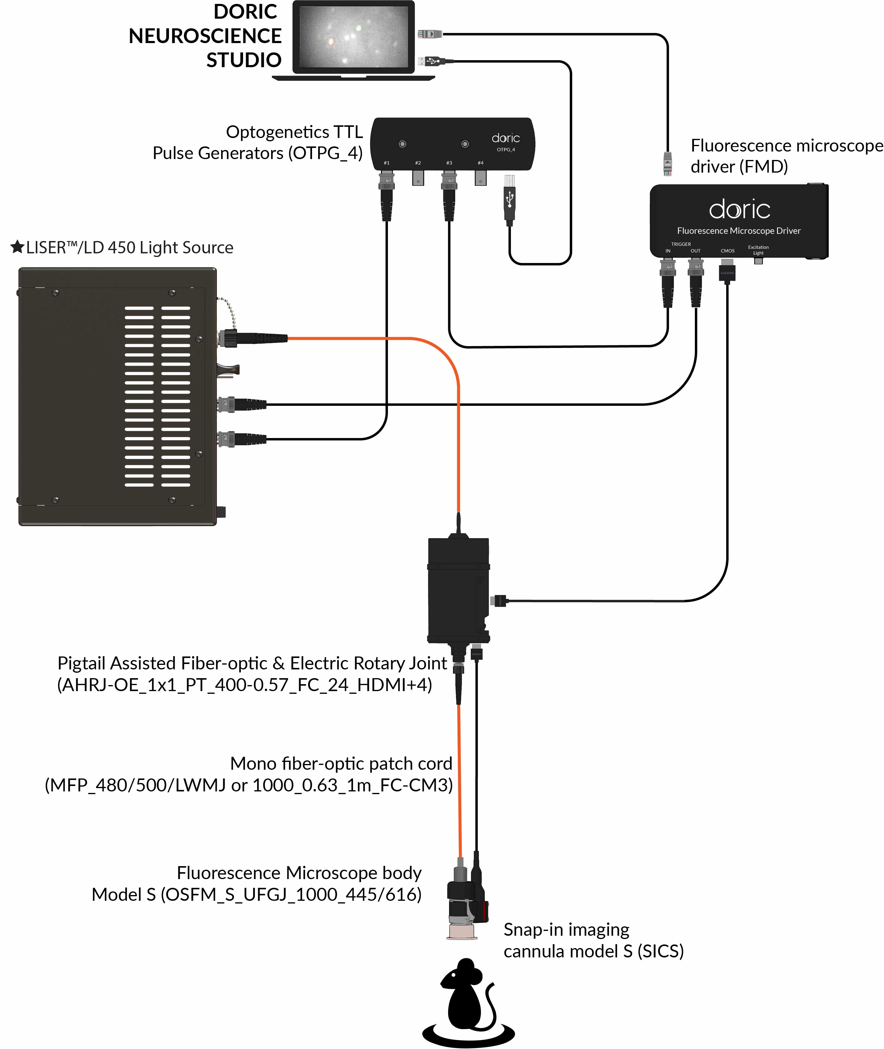
The system includes:

- Snap-in GCaMP/NpHR Fluorescence Microscope Body, Model S, the head mounted microscope
- Fluorescence Microscope Driver, to control the microscope
- ★LISER™ Light Source with 612/69 nm bandpass filter, for GCaMP excitation and Red Opsins activation
- Optogenetics TTL Pulse Generators 4 Channels, for imaging and optogenetic stimulation synchronization
- Optical fiber patch cords, to connect the light source to the microscope
- Fluorescence Microscope Holder
- Clamp for Fluorescence Microscope Holder
- Doric Neuroscience Studio Software
Add optional items:
- Assisted 1x1 Pigtailed Fiber-optic & Electric Rotary Joint - 24 contacts
- Snap-in Microscope Dummy, Model S
- Behavior Camera to record animal behavior
- Workstation
- danse™ analyzing software
Complete your system with:
| Field of View | 700 µm x 700 µm |
| Working distance | 1.1 mm |
| Frame rate | > 45 fps |
| Fluorescence recording | |
|---|---|
| Excitation | 458 / 35 nm |
| Emission | 525 / 40 nm |
| Optogenetic stimulation | |
| Wavelength | 612 / 69 nm |
| Activation intensity | 0 to 55 mW / mm2 |
The Red Fluorescence + Blue Opsins Surface Microscopy System is optimized for RFP-like fluorescence proteins imaging and Blue Opsins (e.g. ChR2) activation.
The system includes:

Add optional items:
Complete your system with:
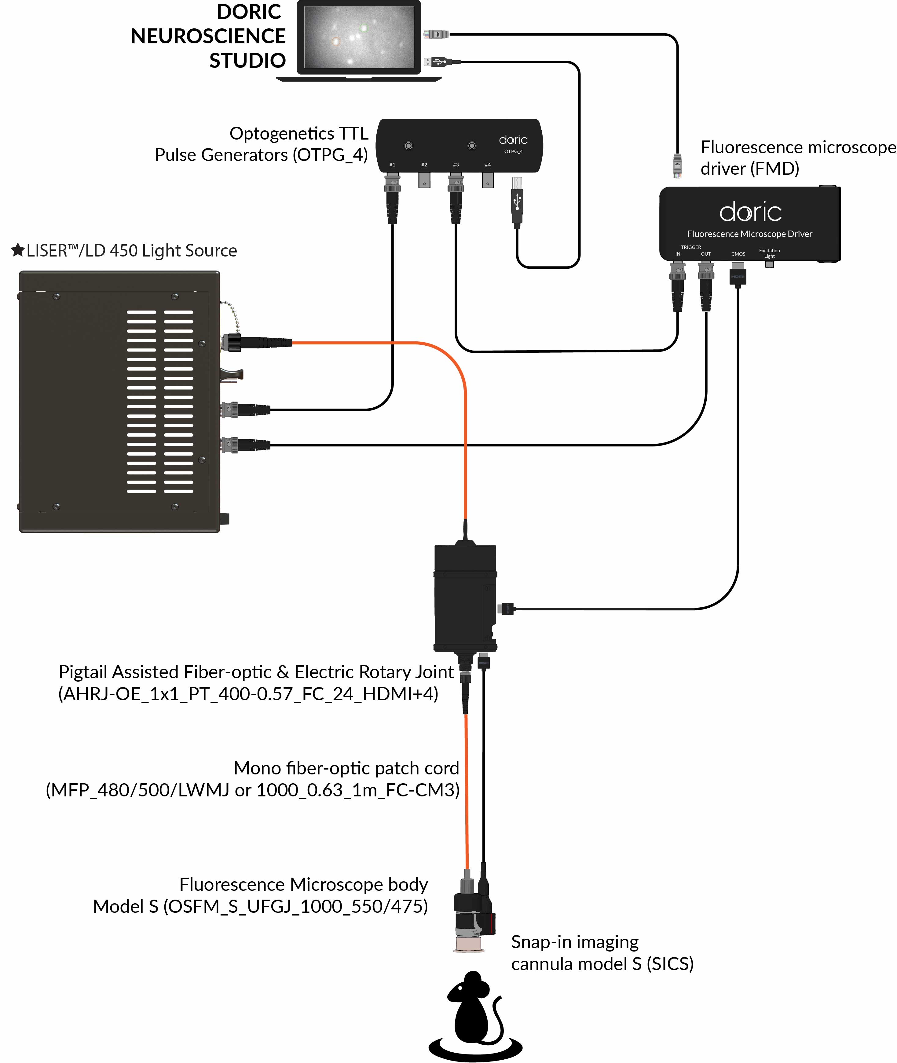
The system includes:

- Snap-in RCaMP/ChR2 Fluorescence Microscope Body, Model S, the head mounted microscope
- Fluorescence Microscope Driver, to control the microscope
- ★LISER™ Light Source with 540/15 nm bandpass filter, for GCaMP excitation and Red Opsins activation
- Optogenetics TTL Pulse Generators 4 Channels, for imaging and optogenetic stimulation synchronization
- Optical fiber patch cords, to connect the light source to the microscope
- Fluorescence Microscope Holder
- Clamp for Fluorescence Microscope Holder
- Doric Neuroscience Studio Software
Add optional items:
- Assisted 1x1 Pigtailed Fiber-optic & Electric Rotary Joint - 24 contacts
- Snap-in Microscope Dummy, Model S
-
Behavior Camera to record animal behavior
- Workstation
- danse™ analyzing software
Complete your system with:
| Field of View | 700 µm x 700 µm |
| Working distance | 1.1 mm |
| Frame rate | > 45 fps |
| Fluorescence recording | |
|---|---|
| Excitation | 540 / 15 nm |
| Emission | 609 / 57 nm |
| Optogenetic stimulation | |
| Wavelength | 465 / 20 nm |
The RCaMP surface microscopy system is optimized for RFP-like fluorescence proteins imaging.
The system includes:

Add optional items:
Complete your system with:
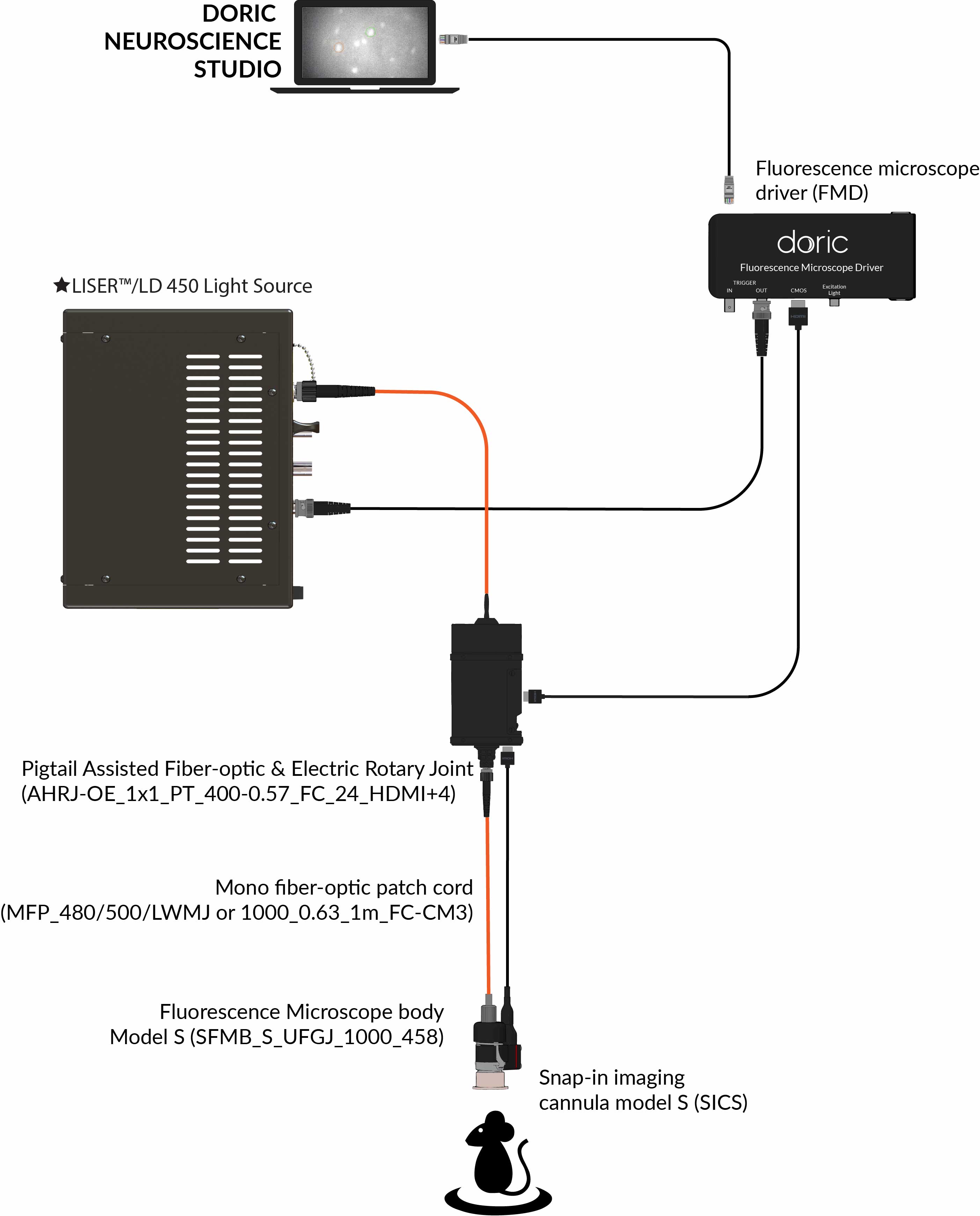
The system includes:

- Snap-in Red Fluorescence Microscope Body, Model S, the head mounted microscope
- Fluorescence Microscope Driver, to control the microscope
- ★LISER™ Light Source with 540/15 nm bandpass filter for Red Fluoescent Proteins excitation
- Optical fiber patch cords, to connect the light source to the microscope
- Fluorescence Microscope Holder
- Clamp for Fluorescence Microscope Holder
- Doric Neuroscience Studio Software
Add optional items:
- Assisted 1x1 Pigtailed Fiber-optic & Electric Rotary Joint - 24 contacts
- Snap-in Microscope Dummy, Model S
- Behavior Camera to record animal behavior
- Workstation
- danse™ analyzing software
Complete your system with:
| Field of View | 700 µm x 700 µm |
| Working distance | 1.1 mm |
| Frame rate | > 45 fps |
| Fluorescence recording | |
|---|---|
| Excitation | 540 / 15 nm |
| Emission | 609 / 57 nm |


