
efocus Fluorescence Microscopy System Gen 3
efocus Fluorescence Microscopy System Gen 3
The 3rd generation efocus Fluorescence Microscope enables calcium imaging with cellular resolution over a larger brain area of freely-moving behaving animals. Its field of view has increased to 650 x 650 µm, and the electronic depth adjustment is 300 µm. A more sensitive sensor of the 3-rd generation microscope requires less fluorescence excitation light, reducing photobleaching and phototoxicity. The microscope body attaches to the imaging cannula with a simple barrel rotation. No tools are required.
The 3rd generation efocus Fluorescence Microscope enables calcium imaging with cellular resolution over a larger brain area of freely-moving behaving animals. Its field of view has increased to 650 x 650 µm, and the electronic depth adjustment is 300 µm. A more sensitive sensor of the 3-rd generation microscope requires less fluorescence excitation light, reducing photobleaching and phototoxicity. The microscope body attaches to the imaging cannula with a simple barrel rotation. No tools are required.
The GCaMP efocus fluorescence microscopy system Gen 3 is optimized for calcium imaging of GFP-like fluorescence proteins.
The system includes:
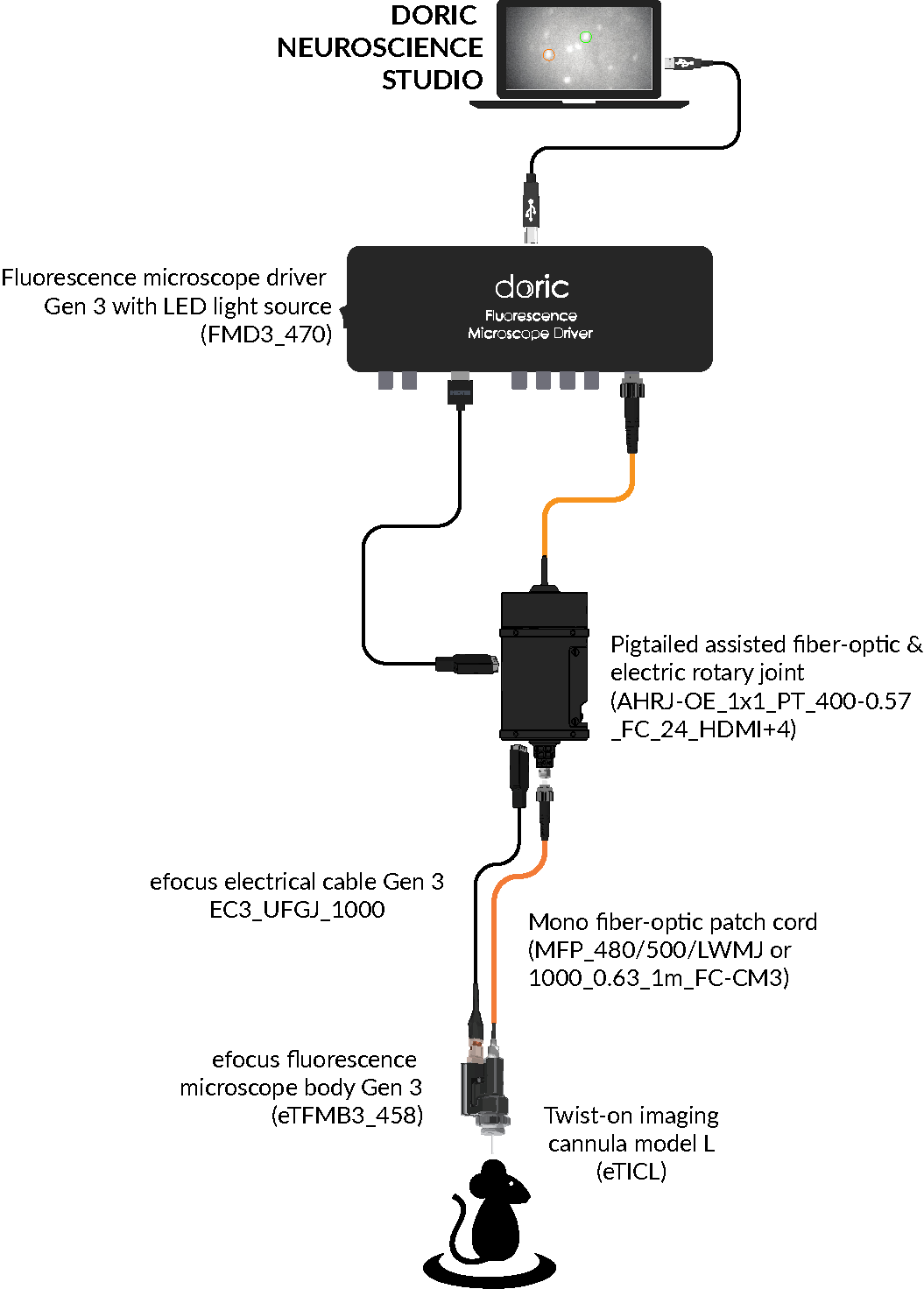
- efocus Green Fluorescence Microscope Body Gen 3, the head mounted microscope
- Fluorescence Microscope Driver Gen 3, to control the microscope
- Electrical cable for efocus Fluorescence Microscope Bodies Gen 3, to connect the microscope to the driver
- Optical fiber patch cords to connect the light source to the microscope
- Fluorescence Microscope Holder 400
- Clamp for Fluorescence Microscope Holder
- Doric Neuroscience Studio Software
Optional items:
- Assisted 1x1 Pigtailed Fiber-optic & Electric Rotary Joint - 24 contacts
- Twist-on Microscope Dummy
- Behavior Camera or CamLoop to record animal behavior
- danse™ analyzing software
Accessories:
| Field of View | 650 µm x 650 µm |
| Working distance adjustment range | 0 - 300 µm |
| Frame rate | up to 45 fps |
| Pc connection | USB 3.0 |
| Computeur requirements | Desktop Computer recommended, Intel Core-i7, 8 GB RAM, Graphic card with 2 GB memory and OpenGL v4.6 compatible |
| Fluorescence recording | |
|---|---|
| Excitation | 458 / 35 nm |
| Emission | 525 / 40 nm |
The RCaMP efocus microscopy system Gen 3 is optimized for calcium imaging Red Fluorescence Proteins.
The system includes:
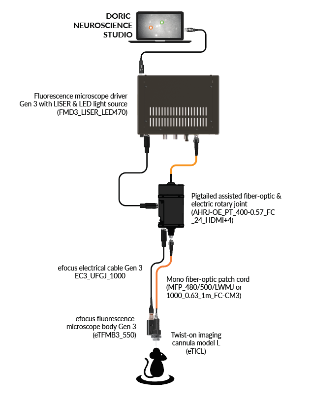
- efocusRed Fluorescence Microscope Body Gen 3, the head mounted microscope
- Fluorescence Microscope Driver with ★LISER™ & LED Light Source with 540/15 nm bandpass filter for Red Fluoescent Proteins excitation
- Electrical cable for efocus Fluorescence Microscope Bodies Gen 3, to connect the microscope to the driver
- Optical fiber patch cords, to connect the light source to the microscope
- Fluorescence Microscope Holder 400
- Clamp for Fluorescence Microscope Holder
- Doric Neuroscience Studio Software
Optional items:
- Assisted 1x1 Pigtailed Fiber-optic & Electric Rotary Joint - 24 contacts
- Twist-on Microscope Dummy
- Behavior Camera or CamLoop to record animal behavior
- danse™ analyzing software
Accessories:
| Fluorescence recording | |
|---|---|
| Field of View | 650 µm x 650 µm |
| Working distance adjustment range | 0 - 300 µm |
| Frame rate | up to 45 fps |
| Pc connection | USB 3.0 |
| Computeur requirements | Desktop Computer recommended, Intel Core-i7, 8 GB RAM, Graphic card with 2 GB memory and OpenGL v4.6 compatible |
| Excitation | 540 / 15 nm |
| Emission | 609 / 57 nm |
The GCaMP + Red Opsins efocus microscopy system Gen 3 is optimized for calcium imaging of GFP-like fluorescence proteins and activation of Red-shifted Opsins (e.g. NpHR3.0, Chrimson, ...).
The system includes: 
- efocus GCaMP/NpHR Fluorescence Microscope Body Gen 3, the head mounted microscope
- Fluorescence Microscope Driver with ★LISER™ & LED Light Source with 612/69 nm bandpass filter, for Red Opsins activation andGCaMP excitation
- Optical fiber patch cords, to connect the light source to the microscope
- Fluorescence Microscope Holder 400
- Doric Neuroscience Studio Software
Optional items:
- Assisted 1x1 Pigtailed Fiber-optic & Electric Rotary Joint - 24 contacts
- Twist-on Microscope Dummy
- Behavior Camera or CamLoop to record animal behavior
- danse™ analyzing software
Accessories:
| Fluorescence recording | |
|---|---|
| Optogenetic stimulation | |
| Field of View | 650 µm x 650 µm |
| Working distance adjustment range | 0 - 300 µm |
| Frame rate | up to 45 fps |
| Pc connection | USB 3.0 |
| Computeur requirements | Desktop Computer recommended, Intel Core-i7, 8 GB RAM, Graphic card with 2 GB memory and OpenGL v4.6 compatible |
| Excitation | 458 / 35 nm |
| Emission | 525 / 40 nm |
| Wavelength | 612 / 69 nm |
| Activation intensity | 0 to 55 mW / mm2 |
The Red Fluorescence + Blue Opsins efocus microscopy system Gen 3 is optimized for calcium imaging of Red Fluorescence Proteins and activation of Blue Opsins (ChR2).
The system includes: 
- efocus RCaMP/CHR2 Fluorescence Microscope Body Gen 3, the head mounted microscope
- Fluorescence Microscope Driver with ★LISER™ & LED Light Source with 540/15 nm bandpass filter for Red Fluorescence Proteins excitation and Blue Opsins (ChR2) activation
- Electrical cable for efocus Fluorescence Microscope Bodies Gen 3, to connect the microscope to the driver
- Optical fiber patch cords, to connect the light source to the microscope
- Fluorescence Microscope Holder 400
- Clamp for Fluorescence Microscope Holder
- Doric Neuroscience Studio Software
Optional items:
- Assisted 1x1 Pigtailed Fiber-optic & Electric Rotary Joint - 24 contacts
- Twist-on Microscope Dummy
- Behavior Camera or CamLoop to record animal behavior
- danse™ analyzing software
Accessories:
| Fluorescence recording | |
|---|---|
| Optogenetic stimulation | |
| Field of View | 650 µm x 650 µm |
| Working distance adjustment range | 0 - 300 µm |
| Frame rate | up to 45 fps |
| Pc connection | USB 3.0 |
| Computeur requirements | Desktop Computer recommended, Intel Core-i7, 8 GB RAM, Graphic card with 2 GB memory and OpenGL v4.6 compatible |
| Excitation | 540/ 15 nm |
| Emission | 609 / 57 nm |
| Wavelength | 465 / 20 nm |
Related Products
-
Choose your option
-
Choose your option

