Opto-Fluidics Solutions
Introduction
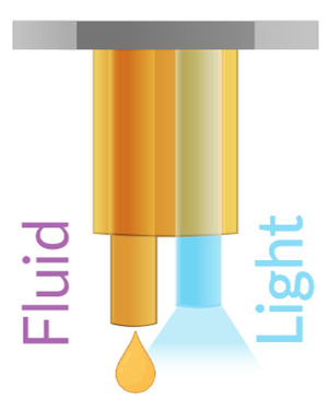
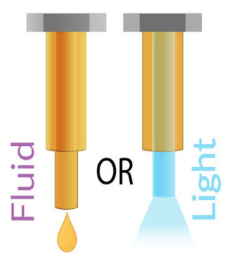
Neuroscience research increasingly leverages advanced tools for manipulating and monitoring neural activity with high precision. Among these, optical fibers and fluid injection systems have emerged as indispensable components in studies involving optogenetics, pharmacological interventions, and neural circuit mapping. Doric Lenses Inc. provides specialized equipment such as the Opto-Fluid Cannula (OmFC, iOFC) that integrates these capabilities, offering researchers versatile and modular solutions.
Optical fiber in Neuroscience Optical fibers are used in neuroscience for delivering and/or collecting light to specific brain regions. The used of optical fibers give access to manipulate or record deep brain regions with high specificity on the neuron population due to the genetics modification, and compatible with in-vivo and freely-moving experiments.
Fluid Injection in Neuroscience Controlled fluid injection into brain tissue allows researchers to deliver pharmacological agents, viral vectors, or other solutions with spatial and temporal specificity. This is essential for:
-
Neuropharmacology: Studying the effects of drugs on specific brain circuits.
-
Viral Delivery: Introducing genetic constructs for functional studies.
-
Lesion Studies: Creating localized lesions to investigate brain function.
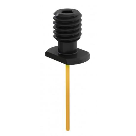
Figure 1. Example of an opto-fluid cannula setup integrating optical fiber and fluid delivery.
Opto-Fluid Cannulas
Doric Lenses offers solutions that combine optical stimulation and fluid injection capabilities in a single implantable device. These are divided in 3 main categories :
-
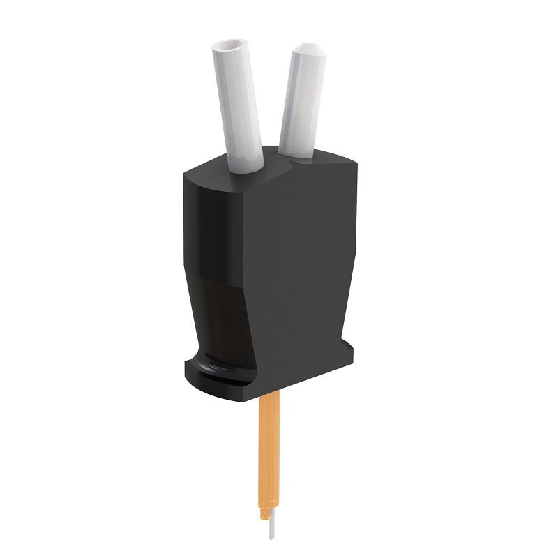
Multiple Fluid Injections Cannulas
-
Designed with chronically implanted optical fiber combined with fluid guiding tube for repeated fluid delivery to the same brain region removable using micro-injectors.
-
Enables chronic experiments and longitudinal studies.
-
Features precise alignment between optical and fluid delivery pathways.
-
-
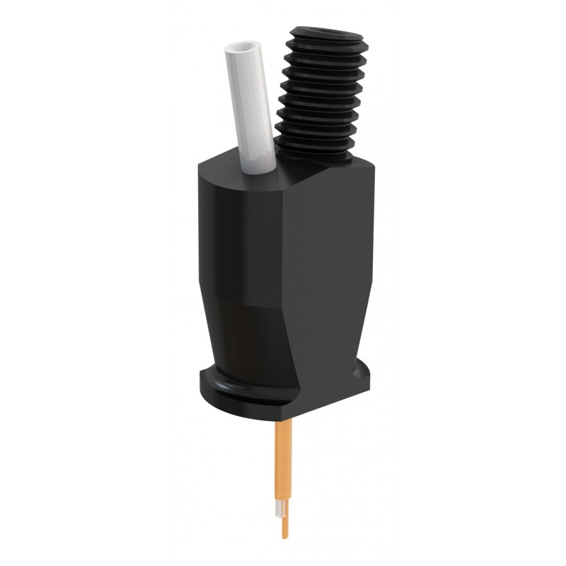
Opto-Fluid Cannula with Interchangeable Injectors
-
Provides modularity through swappable injectors (optics vs fluid) with smaller implant dimensions.
-
Allows flexibility in experimental protocols.
-
Maintains consistent alignment and sealing for reliable injections.
-
-
Opto-Fluid Cannula with Single-shot Injectors
-
-
Compare table part 1.
| Image | Image tip | Compatible Stereotaxic Holder | Compatible Fluid Injector | Compatible Optical Injector | |
|---|---|---|---|---|---|
| OsFC | 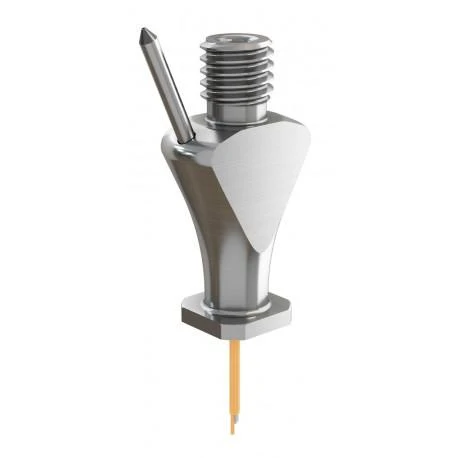 |
 |
SCH_OmFC_ZF  |
Single injection, should be pre-filled | Use patchcord with CM3 termination |
|
OmFC SM3 |
 |
|
SCH_OmFC_SM3 |
OI_iOFC-M3 |
|
|
OmFC ZF1.25 |
 |
SCH_OmFC_ZF |
FI_OmFC_ZF |
Use patchcord with ZF1.25(F) termination | |
| iOFC M3 |
 |
 |
FCA_1.25_SM3 |
FI_iOFC-M3 |
OI_iOFC-M3 |
Example Research Protocols
-
Optogenetic Manipulation of Fear Circuits
-
Use the OmFC to deliver a virus encoding channelrhodopsin into the basolateral amygdala.
-
Two weeks later, use the optical fiber to deliver blue light stimulation during fear conditioning.
-
Citation: Johansen, J. P., et al. (2010). Optical activation of lateral amygdala pyramidal cells instructs associative fear learning. PNAS, 107(28), 12692-12697. https://doi.org/10.1073/pnas.1002418107
-
-
Drug Microinjection and Behavioral Assessment
-
Inject muscimol (a GABA agonist) into the prefrontal cortex using a Doric opto-fluid cannula.
-
Record changes in working memory task performance.
-
Citation: Preston, A. R., & Eichenbaum, H. (2013). Interplay of hippocampus and prefrontal cortex in memory. Current Biology, 23(17), R764-R773. https://doi.org/10.1016/j.cub.2013.05.041
-
-
Viral Delivery and Optogenetic Inhibition in Cortical Circuits
-
Use interchangeable injectors to deliver AAV-Cre-dependent halorhodopsin into the medial prefrontal cortex of Cre-expressing mice.
-
Inhibit activity during decision-making tasks.
-
Citation: Kim, C. K., et al. (2016). Simultaneous fast measurement of circuit dynamics at multiple sites across the mammalian brain. Nature Methods, 13(4), 325-328. https://doi.org/10.1038/nmeth.3770
-
Limitations and Technical Considerations While opto-fluid cannulas offer powerful experimental capabilities, several limitations and best practices should be considered:
-
Optical Fiber Size and Tissue Damage: Larger core fibers or those with rigid ferrules can cause mechanical damage and gliosis around the implantation site, potentially affecting neural activity and experimental outcomes. Using thinner, low-profile fibers helps minimize these effects but may reduce light transmission efficiency.
-
Optimal Fluid Delivery Rates: Injection rates that are too fast can cause backflow or tissue disruption, while very slow rates may be ineffective. A commonly recommended infusion rate is between 100–500 nL/min, depending on the viscosity of the solution and the target brain region (Myers, 1966; Veenman et al., 1992).
-
Long Tubing Considerations: Long and flexible tubing can introduce dead volume and delay between the start of injection and fluid delivery, which complicates time-sensitive protocols. Pre-filling tubing and validating flow rate are essential steps in reducing variability.
-
Clogging of Injectors: Injectors may clog over time due to particulates in solutions or dried residues. To prevent clogging, researchers should filter all solutions, flush injectors after use with sterile saline or appropriate solvents, and avoid prolonged storage with fluid in the lines.
Applications in Research These integrated tools support diverse research paradigms, including:
-
Behavioral Neuroscience: Correlating light-evoked activity with behavior.
-
Circuit Dissection: Combining optogenetics with drug injections to parse network functions.
-
Therapeutic Studies: Assessing effects of targeted delivery of neurotherapeutics.
-
Longitudinal Studies: Monitoring chronic effects of repeated interventions with high spatial accuracy.
External References
|
1. Separate anterior paraventricular thalamus projections differentially regulate sensory and affective aspects of pain. |
|
1. Noemi Rook et al. AAV1 is the optimal viral vector for optogenetic experiments in pigeons (Columba livia). Commun Biol.4, 100 (2021). Brain region: entopallium (important primary visual area in the pigeon) |
| 2. Fernández-García et al. M2 cortex-dorsolateral striatum stimulation reverses motor symptoms and synaptic deficits in Huntington’s disease. eLife 9, e57017 (2020). Brain region: cortico-striatal (M2-DLS) |
| 3. Brendan D. Hare et al. Optogenetic stimulation of medial prefrontal cortex Drd1 neurons produces rapid and long-lasting antidepressant effects. Nature Communications 10, (2019). Brain region: medial prefrontal cortex (mPFC) |
|
4. Michelle M. Sidor et al. In vivo Optogenetic Stimulation of the Rodent Central Nervous System. J. Vis. Exp.95, e51483 (2015). Brain region: ventral tegmental area |
|
5. Gradinaru V et al.Targeting and readout strategies for fast optical neural control in vitro and in vivo. J Neurosci.27, 14231-8 (2007). |
| 6. addgene Optogenetics guide |
| 7. Deisseroth lab Optogenetics Ressource |