
Snap-in Imaging Cannula Model L
Snap-in Imaging Cannula Model L
For deep brain regions use the Snap-in Imaging Cannula Model L that brings the image from inside the brain to the skull surface with an image guiding gradient-index (GRIN) lens. Snap-in Imaging Cannulas Model L are compatible with:
- Snap-in 1-color Fluorescence Microscope Bodies Model L (SFMB, OSFM)
- 2-color Fluorescence Microscope Bodies model L (2CFM)
…generating
For deep brain regions use the Snap-in Imaging Cannula Model L that brings the image from inside the brain to the skull surface with an image guiding gradient-index (GRIN) lens. Snap-in Imaging Cannulas Model L are compatible with:
- Snap-in 1-color Fluorescence Microscope Bodies Model L (SFMB, OSFM)
- 2-color Fluorescence Microscope Bodies model L (2CFM)
Three different depth ranges of brain tissue are accessed with different lens lengths while fine adjustment is done with the protrusion adjustment rings that come with each cannula. It is recommended to use the smallest lens for the desired depth for a higher stability and image quality. The standard working distance of the imaging cannulas (distance between the tip of the GRIN lens and the imaged plane) is 80 µm. Custom working distance can be provided upon request.
Prism tip is available to image a transversal section of the brain (prism orientation on option):
|
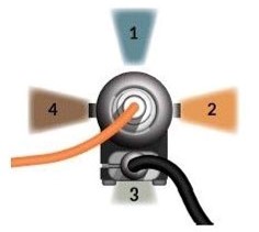
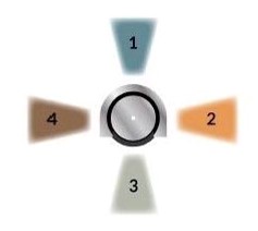
Microscope Body top view (for reference) |
Imaging Cannula top view (for reference) |
 |
|
Notes
- Each Snap-in Imaging Cannula Model L is provided with one of each of the five models of Protrusion Adjustment Rings Model L. If more rings are needed, a set can be purchased separately (PARS_L).
- 500 microns diameter GRIN lenses with prism are now protected with a metallic tubing (outer diameter 750 um). Imaging cannula with prism tip are still available without the metallic protection on custom request (contact sales@doriclenses.com)
| Penetration depth | - Type D: 0 - 3.4 mm - Type V: 2.9 - 5.9 mm - Type E: 5.4 - 8.3 mm |
| GRIN lens numerical aperture | 0.5 |
| Working distance | 80 µm |
Related products
-
Choose your option
-
Choose your option
-
Choose your option


