"NEW" Bundle-imaging Fiber Photometry System


-
Doric's Bundle-imaging Fiber Photometry System (BFMC) is fully integrated, offering a hassle-free, plug-and-play solution. To record green and red photometry signals, the BFMC uses built-in LEDs to illuminate the entire sample port, and a CMOS sensor to simultaneously image the fluorescent signal from every fiber within the bundle. The fluorescent light collected from each fiber within the bundle creates circular spots on a CMOS sensor. The electrical read-out from pixels within each fiber image correlates with the calcium activity of the corresponding brain site.
The BFMC system is equipped with 8 Digital Input/Output (DIO) ports and a HD25 adapter for synchronization and closed-loop experiments with external devices such as behavior cameras, operant conditioning chambers, video tracking software, and optogenetic light sources.
To combine bundle photometry with optogenetics see Doric BFTO systems.
-
The 1-colors Bundle-imaging Fiber Photometry System records activity-independent green indicator (i.e. GCaMP) fluorescence, excited by 405, 415, 425, or 440 nm light (isosbestic point), and its activity-dependent fluorescence, excited by 470 nm light, from several fiber-connected brain sites with a CMOS image sensor. The sites labelled with green indicators can be on the same animal or distributed over many animals. The respective excitations are interleaved or sequentially applied to separate activity-dependent and activity-independent fluorescence emissions.
The system includes: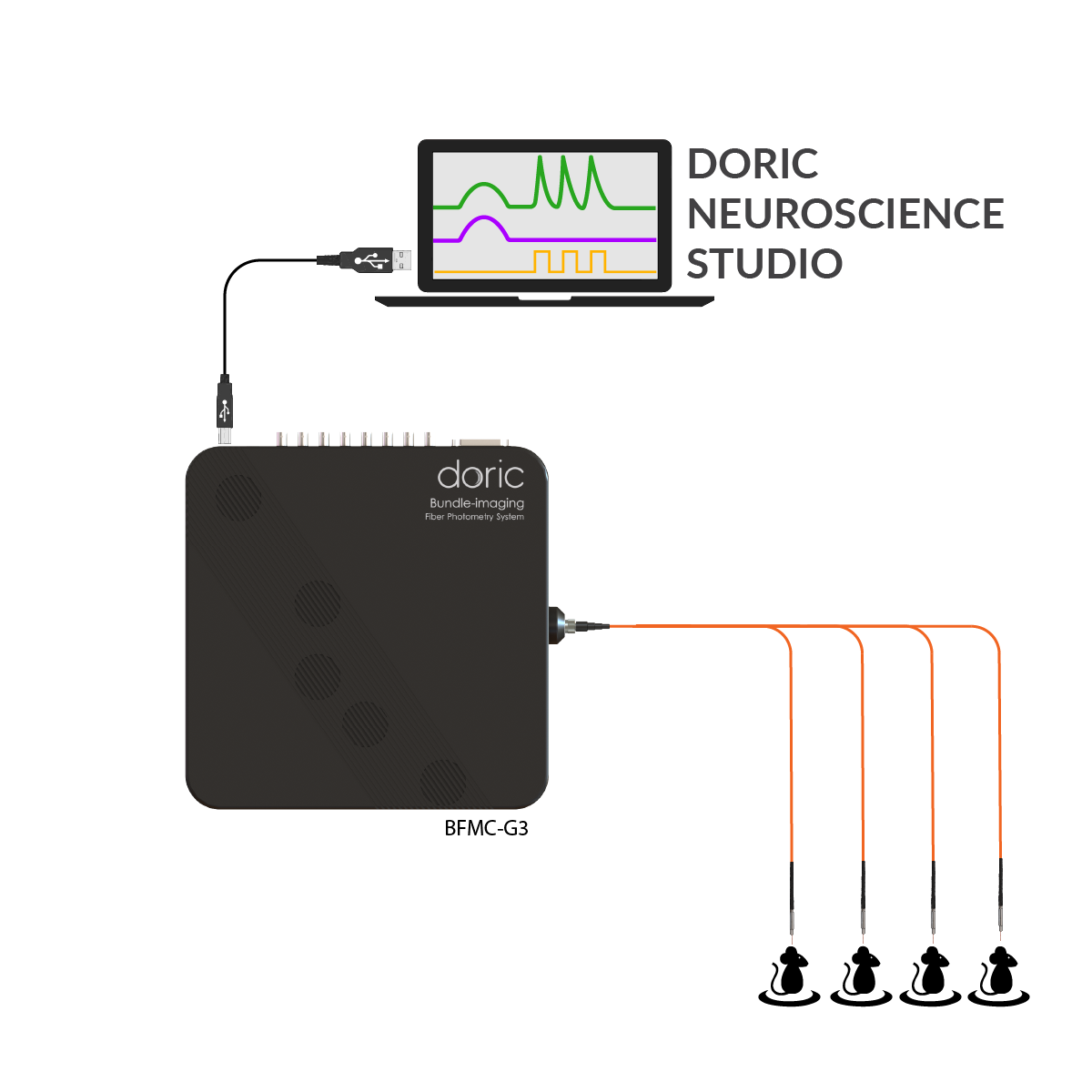
- Bundle-imaging Fiber Photometry System
- All required electrical cables and patch cord
Optional items:- Fiber Photometry Cannula Holder to record during surgery
- Optical Power meter to monitor and calibrate excitation power
- Behavior Camera or CamLoop to record animal behavior
- Workstation
- danse™ analyzing software
- Pigtailed 1x1 Fiber-optic Rotary Joint with Holder (fixed or gimbal) for each animal in a separate cage with a single brain site
Accessories:- Low-autofluorescence Branching Fiber Patch cords to bring light to the animal
- High-density Fiber-optic cannula and patch cord for multiple fibers with high placing precision chronic implant
- Fiber-optic Cannulas for chronic implant
- Mating sleeves for ferrule to ferrule interconnect
- Bundle-imaging Fiber Photometry System
-
The 2-colors Bundle-imaging Fiber Photometry System records activity-independent and activity-dependent fluorescence from a green indicator (i.e. GCaMP) and red fluorophore (reference or indicator). The signals are collected from several fiber-connected brain sites with two CMOS image sensors for the green and red channels. The excitation wavelength for the isosbestic reference can be selected between 405, 415, 425, or 440 nm, while the excitation wavelengths are 470 nm for the green channel and 560 nm for the red channel. The sites labelled with fluorophores can be on one animal or distributed over many animals. The respective fluorophore excitations are interleaved or sequentially applied to separate different fluorescence emissions.
The system includes:- Bundle-imaging Fiber Photometry System
- All required electrical cables and patch cord
Optional items:- Fiber Photometry Cannula Holder to record during surgery
- Optical Power meter to monitor and calibrate excitation power
- Behavior Camera or CamLoop to record animal behavior
- Workstation
- danse™ analyzing software
- Pigtailed 1x1 Fiber-optic Rotary Joint with Holder (fixed or gimbal) for each animal in a separate cage with a single brain site
Accessories:- Low-autofluorescence Branching Fiber Patch cords to bring light to the animal
- High-density Fiber-optic cannula and patch cord for multiple fibers with high placing precision chronic implant
- Fiber-optic Cannulas for chronic implant
- Mating sleeves for ferrule to ferrule interconnect
- Bundle-imaging Fiber Photometry System
-
Mini Cube: Wavelength range 350 to 1100 nm Field of view Ø2.5 mm Objective NA 0.40 Maximum number of sites
* could be limited by patchcord manufacturing- 20x core 400 µm NA0.37
- 60x core 200 µm NA0.37
- 100x core 100 µm NA0.37Excitation Uniformity 10% over FOV Optical fiber compatibility Core diameter 100, 200 or 400 µm NA 0.37 Optical filter attenuation OD 5 outside band Optical fiber connector SMA 1-color (GCaMP) Spectral bandwidth - Excitation #1 : 400-410, 410-420, 421-433, or 433-445 nm
- Excitation #2 : 460-490 nm
- Emission : 500-550 nm
2-colors (GCaMP + RCaMP) Spectral bandwidth - Excitation #1 :400-410, 410-420, 421-433, or 433-445 nm
- Excitation #2 : 460-490 nm
- Excitation #3 : 555-570 nm
- Emission #1 : 500-540 nm
- Emission #2 : 580-680 nm
Sensor Type CMOS Image sensor Quantum Efficiency 82% at 520 nm Resolution 1024 x 1024 px Pixel Size 5.86 µm x 5.86 µm Frame rate up to 60 Hz Build-in LEDs Max Current
500 mA Max Output Power
(fibre 400 µm, NA0.37)
LED 400-410 nm = 700 µW (at 200 mA, CW)
LED 460-490 nm = 700 µW (at 200 mA, CW)
LED 555-570 nm = 130 µW (at 200 mA, CW)DI/O
Number of port 32 Maximum sampling rate 10 kSps Maximum output frequency - 8 BNC I/O,
- 1 DB25 with 8 Input/Output, 8 Input, and 8 Output
(DB25 adapter to wires included)Computer Requirement
Operating System
Microsoft Windows 10; 64-bit Memory 8 GB RAM minimum
16 GB RAM recommendedProcessor speed 3 GHz and 8 cores Hard Drive 500 MB of free hard disk space>
* SSD recommended
Connection to computer USB-3 (cable included) Physical properties Size
310x310x66mm -
Drawing Bundle-imaging Fiber Photometry System Gen 3
User ManualFrEn Bundle-imaging Fiber Photometry System Gen3
- le choix d'une sélection entraîne une actualisation complète de la page