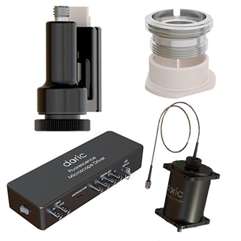
Miniaturized Fluorescence Microscopy
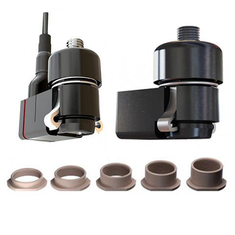
Miniature Fluorescence Microscope monitors the activity of neural circuitry in freely moving animals (e.g., mice or rats) with cellular resolution. It can be combined with optogenetic activity modulation within or outside its field of view.
1-color Deep Brain Fluorescence Microscope uses gradient-index relay lens to re-image fluorescence flashes of neuronal populations found as deep as 8 mm into the brain.
1-color Surface Fluorescence Microscope uses an aspheric lens to look up to 150 µm deep into the brain with a larger field of view and better imaging quality.
2-color Fluorescence Microscope enables simultaneous imaging of GFP- and RFP-like fluorophores in deep-brain or surface configuration.