Optogenetics Solutions
Optogenetics is a groundbreaking technique that provides real-time, optical control of neuronal activity. Throughout the years, it has played a vital role in dissecting brain circuitry, probing the neural basis of behavior, modeling neurological disorders, and developing next-generation therapies in neuroscience and psychiatry.
The process requires expressing opsins, light-sensitive ion channels or pumps, in a specific neuronal population. For chronic experiments, a fiber-optic cannula is then surgically implanted above the target region to deliver a specific wavelength of light, activating or inhibiting the opsin-expressing cells. For example, blue light activates opsins like Channelrhodopsin (ChR2), while yellow or red light targets opsins such as NpHR or Jaws. For a list of available opsins, see the OSPIN APPLICATION NOTE.

At Doric Lenses, we offer a comprehensive range of light sources for optogenetics, all developed and manufactured in-house, including LED modules, laser diodes, and LISERTM. In addition, we provide a complementary ecosystem of optical components such as light splitters, combiners, attenuators, optical rotary joints, patch cords, and cannulas—including components that combine optogenetics with other types of measures (behavior, fiber photometry, Doric Miniscope, fiber photometry, etc.).
This page outlines the key features of Doric light sources. However, we highly recommend consulting with specialists to select the most suitable option for your specific application.
Light Source
Choosing the right light source is one of the most important steps when designing an optogenetics experiment. The optimal light sources depend on opsin requirements (excitation wavelength & intensity), the size of the region(s)-of-interest and whether the animal is freely moving.

LED Light Sources
LEDs are among the most commonly used light sources in optogenetics, and are particularly suitable for experiments stimulating large regions-of-interest (with 400 µm - 1mm diameter fibers) at moderate light intensities. Their large emitter, inherent safety, reliability, and cost-effectiveness make them a preferred option across a variety of experimental designs.
At Doric Lenses, we offer a wide range of CLED wavelengths in the near-UV (350-400 nm), visible (400-700 nm) and near IR range (700-1100 nm) for standard optogenetic applications.
-
Connectorized LED + Rotary Joint
-
Wireless Optogenetics Headstage
Laser Diode
Laser diodes are high-powered light sources with small emitter ideal for optogenetic experiments requiring high intensity of light in small regions-of-interest (< 200um). Their narrow beam profile and monochromatic light output make lasers ideal for illumination through small-diameter patch cords (typically 50–200 µm) and are a great solution when precise spatial targeting is required.
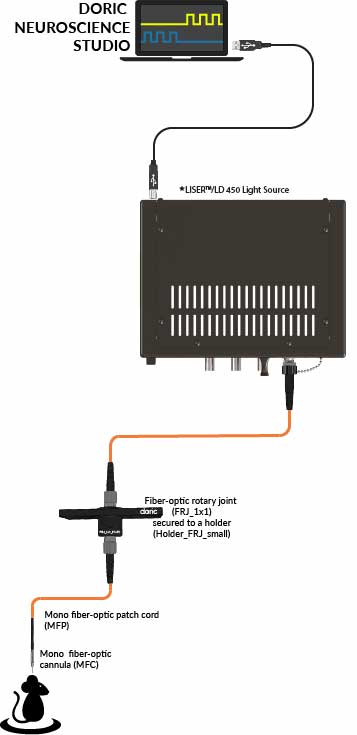
LISER™
LISER™ (Laser-Induced Spontaneous Emission of Radiation) is an advanced light source providing high-intensity, broad-spectrum illumination.
The system uses a laser pumped Ce:YAG (cerium-doped yttrium aluminum garnet) crystal, which emits a continuous spectrum from yellow to red (500–650 nm). This configuration provides exceptionally high optical power (> 85mW full spectrum in a 200 um NA 0.57 fiber) in a spectral range that is difficult to achieve with standard LEDs or laser diodes, particularly around 590 nm, optimal for activating inhibitory opsins such as NpHR and Jaws.
In addition to its broad-spectrum output, LISER™ includes an integrated blue excitation source (laser or LED), making it a versatile, all-in-one solution capable of supporting both excitatory and inhibitory optogenetics simultaneously.
The LISER also includes its own internal drivers, allowing independent control of two wavelength channels in the same brain region (e.g. 450 nm + 590 nm).
Key Features:
-
Full-Spectrum Coverage: Simultaneously delivers blue excitation plus tunable yellow-red wavelengths using interchangeable optical filters (e.g., 525, 559, 582, 593, 612 nm).
-
High Output Power: Ideal for experiments requiring strong illumination intensity, including bilateral and large-area stimulation.
-
Supports Large Core Patch Cords for wide brain region illumination: Optimized for 200 - 400 µm core diameters, enabling uniform wide-area stimulation.
Light Source Driver
To operate Doric LED and Laser diode modules, a dedicated light source driver is required. The driver can be operated in 1) standalone, 2) external modes (Analog or TTL), or 3) program sequences using free Doric Neuroscience Studio (DNS) software. In DNS, users can define a wide range of light stimulation patterns including continuous wave output, square pulse trains, sine waveforms, and more complex waveforms from text file input.
Watch the tutorial video for step-by-step guidance on configuring LED drivers.The drivers are available in 1-4 channel configurations, allowing simultaneous control of up to four LED or laser modules independently.
Doric offers two configurations for drivers:
-
Integrated Driver Units:
In this setup, each driver channel has a built-in LED or laser, offering a compact and user-friendly solution.
-
Modular Driver Setup:
In this version, the LED/laser remains separate from the driver. This provides flexibility to switch between different LED or laser modules using a single driver.
Note: LED drivers are only compatible with LEDs, and LD drivers only with lasers.
Light Splitters
Some neuroscience experiments, such as bilateral, multi-site and/or multi-animal optogenetics, require splitting the light from a single light source into multiple fibers. To effectively split light for these applications, several solutions are available. The optimal splitter type depends on the light source (LED, LISER or laser diode), the intensity requirements and the size of the region-of-interest.

Available Splitter Options:
-
Doric Mini Cube (DMC)- Intensity Division
-
Splitter Branching Fiber-optic Patch Cords
-
1x2 Fiber-optic Rotary Joints - Intensity Division
Doric Mini Cube (DMC)
The DMC contains dichroic mirrors that evenly divide the intensity of light from one light source into multiple fibers (2-4 sites). Note that DMC has a built-in numerical aperture (NA) of 0.3, which limits the effective power transmission when used with patch cords of higher NA. Thus, the DMC is ideal for laser diodes, but less optimized for CLED and LISER.
The output transmission of DMC can be 20-40% transmission, depending on the number of outputs (2-4)
Fiber-optic Rotary Joint Splitter (FRJ)
Fiber-optic Rotary Joint Splitter is an advanced version of DMC light splitter, which is integrated in a rotary joint for bilateral or dual-site optogenetics in freely-moving animals. It maintains comparable optical efficiency to the DMC (~40% per output channel), while enabling rotational freedom during behavior tasks. Currently, only the 1×2 configuration is offered.
FRJ has a built-in numerical aperture (NA) of 0.22, which makes it more ideal for laser diodes, but less optimized for CLED and LISER.
Splitter Patch Cords
The Splitter Branching Fiber-optic Patch Cord (SBP) evenly splits the light from a large-core optic fiber into 2 or more small-core fibers. The output transmission for each branch depends on the surface area difference between the large and small cores (generally ~16% transmission*). Splitter Patch Cords are well-suited for light sources with large emitters, such as LEDs and LISER. However, when used with laser sources (which have very small emitters), the output power is much lower than with DMC or FRJ splitter.
Light Combiners
Certain experiments require delivering multiple wavelengths of light to the same target brain region for various applications. For instance, to manipulate different neural populations expressing different opsins (ChR2 & ChrimonR), for bidirectional control of the same population (excitation/inhibition). Some special opsins are engineered to respond to mutliple wavelengths, such as step-function opsins, or can be toggled on/off with distinct wavelengths.
Doric Lenses offers several reliable and customizable light-combining solutions:
-
LISERTM
-
Combined LEDs (LEDC)
-
Combined LEDs Fiber Rotary Joint (LEDFRJ)
-
Doric Mini Cube (DMC) -intensity division

LISER™ Multi-Wavelength Light Source
One of the most advanced options is the LISER™, which integrates a blue light source (either LED or laser) along with a Ce:YAG-based broadband emitter that covers the yellow to red spectrum to a single optical path. The emission can be filtered to narrow down the wavelength range (e.g., 525, 590, 612 nm), and both the blue and filtered yellow/red excitations. This all-in-one device includes its own internal drivers, allowing independent control of two wavelength channels (e.g. 450 nm + 590 nm).
Combined LEDs
Another compact solution is the Combined LEDs, which uses dichroic mirrors to merge up to four integrated LEDs into a single output connector. Each LED channel can be independently controlled using Doric LED drivers.
The Combined LED box is also available with an integrated fiber-optic rotary joint (Combined LEDs with Fiber-optic Rotary Joint), ideal for multi-color stimulation in freely moving animals. Not only does this solution reduce wire tangling, but it also eliminates optical losses typically introduced by adding external rotary joints, making them particularly suitable for low-power LEDs like 595 nm.
Doric Mini Cube (DMC) - Wavelength Division
The DMC with wavelength division dichroic mirrors also serves as a robust and flexible light combiner. It accommodates up to four input channels of different wavelengths and works just as well to combine and/or split different wavelengths.
Light Attenuators
Maintaining optogenetics light power within the appropriate range is essential to prevent issues such as photobleaching, tissue overheating, and other unwanted side effects. However, under certain conditions, the light output from the source may exceed the optimal level for optogenetic applications—even when operating close to the threshold current setting. It is also not recommended to drive a light source close to the threshold for stability issues. In such cases, an additional component is required to deliberately reduce the light intensity.
Doric Lenses offers different customizable light-attenuating solutions:

U-bracket
U-bracket is a simple optic cube which contains an inter-changeable neutral density filter to attenuate light, or a spectral filter to reduce the spectral range of a light beam. The attenuation and spectral filter can be customized by the user.
Attenuating Mono Fiber-optic Patch Cords
Fiber-optic patch cords with an integrated attenuating filter are ideal for applications where optical power coupled into a fiber is too high, i.e. fiber photometry excitation. Addition of attenuating filter does not affect light distribution inside the optical fiber, only transmission is reduced. Different optical fibers or attenuating factors are possible.
Notes:
-
For attenuating patch cords, optical transmission is specified for visible light and measured at a wavelength of 465 nm. At shorter wavelengths—such as 405 nm (UV)—the transmission is typically about half the specified value.
-
Attenuating filters can also be integrated into branching fiber-optic patch cords upon request.
External References
|
1. Separate anterior paraventricular thalamus projections differentially regulate sensory and affective aspects of pain. |
|
1. Noemi Rook et al. AAV1 is the optimal viral vector for optogenetic experiments in pigeons (Columba livia). Commun Biol.4, 100 (2021). Brain region: entopallium (important primary visual area in the pigeon) |
| 2. Fernández-García et al. M2 cortex-dorsolateral striatum stimulation reverses motor symptoms and synaptic deficits in Huntington’s disease. eLife 9, e57017 (2020). Brain region: cortico-striatal (M2-DLS) |
| 3. Brendan D. Hare et al. Optogenetic stimulation of medial prefrontal cortex Drd1 neurons produces rapid and long-lasting antidepressant effects. Nature Communications 10, (2019). Brain region: medial prefrontal cortex (mPFC) |
|
4. Michelle M. Sidor et al. In vivo Optogenetic Stimulation of the Rodent Central Nervous System. J. Vis. Exp.95, e51483 (2015). Brain region: ventral tegmental area |
|
5. Gradinaru V et al.Targeting and readout strategies for fast optical neural control in vitro and in vivo. J Neurosci.27, 14231-8 (2007). |
| 6. addgene Optogenetics guide |
| 7. Deisseroth lab Optogenetics Ressource |